
BIOL-380 · Lesson 4 of 12 · 29 min
Antigen presentation and the MHC
Antibodies recognize the intact shapes of molecules, but T cells cannot. A T cell receptor recognizes only a short peptide fragment of a protein, held in the groove of a host molecule called a major histocompatibility complex (MHC) molecule on the surface of another cell. This arrangement, discovered in the 1970s, solves a basic problem: it lets T cells inspect the contents of other cells, including proteins made by viruses hidden inside them. This lesson describes the two classes of MHC molecule, the pathways that load them with peptides, the special role of dendritic cells, the extraordinary genetic diversity of the human MHC and its consequences for transplantation and disease.
The discovery of MHC restriction
The MHC was first identified not by immunologists studying infection but by geneticists studying the rejection of tumor and skin grafts between strains of mice. In the 1930s and 1940s Peter Gorer and George Snell showed that the fate of grafts depended largely on a single genetic region, which Snell called the histocompatibility-2 (H-2) locus; Jean Dausset found the corresponding human antigens on white blood cells in 1958, naming them human leukocyte antigens (HLA). Baruj Benacerraf then showed that genes in the same region controlled whether animals could make immune responses to particular antigens. Snell, Dausset and Benacerraf shared the 1980 Nobel Prize.
Why should transplantation antigens control immune responses? The answer came in 1974 from Rolf Zinkernagel and Peter Doherty, working at the Australian National University. They found that cytotoxic T cells from mice infected with lymphocytic choriomeningitis virus killed virus-infected target cells only if the targets shared MHC molecules with the mouse the T cells came from. T cells therefore recognized not virus alone, but virus together with self MHC, a phenomenon called MHC restriction. Zinkernagel and Doherty received the Nobel Prize in 1996. In 1987 Pamela Bjorkman, Don Wiley and Jack Strominger solved the crystal structure of the human class I molecule HLA-A2 and found a peptide bound in a groove on its top surface, showing exactly what T cells see.
The two classes of MHC molecule
MHC class I molecules consist of a membrane-anchored heavy chain, with three domains called α1, α2 and α3, noncovalently associated with a small protein, β2-microglobulin, which is encoded outside the MHC. The α1 and α2 domains form a platform of eight β strands topped by two α helices, with a groove between them that binds peptides of usually 8 to 10 amino acids. The groove is closed at both ends, so the peptide's ends are tucked into pockets, and two or three of its side chains, called anchor residues, fit into specific pockets in the floor of the groove. Each MHC variant prefers peptides with particular anchor residues, its binding motif. Class I molecules are found on almost all nucleated cells and present peptides to CD8 T cells; the CD8 coreceptor binds the α3 domain.
MHC class II molecules consist of two membrane-anchored chains, α and β, each contributing one domain to the groove. The class II groove is open at both ends, so it binds longer peptides of about 13 to 25 amino acids, with a core of nine residues in the groove and the ends hanging out. Class II molecules are found mainly on professional antigen-presenting cells, dendritic cells, macrophages and B cells, and on thymic epithelium, although interferon-γ can induce them on other cells. They present peptides to CD4 T cells; CD4 binds the β2 domain. The restriction of class II to specialized cells means that CD4 T cells, which orchestrate responses, are activated only by cells whose job is to report on infection.
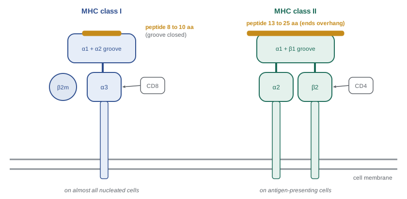
| Feature | MHC class I | MHC class II |
|---|---|---|
| Chains | Heavy chain (α1–α3) plus β2-microglobulin | α and β chains |
| Peptide length | Usually 8–10 amino acids; groove closed | About 13–25 amino acids; groove open |
| Cells expressing | Almost all nucleated cells | Dendritic cells, macrophages, B cells, thymic epithelium (inducible on others) |
| Source of peptides | Proteins in the cytosol and nucleus (endogenous) | Proteins in endosomes (exogenous or engulfed) |
| Where peptides load | Endoplasmic reticulum | Late endosomes (MIIC compartment) |
| T cells recognizing | CD8 T cells | CD4 T cells |
| Human genes | HLA-A, HLA-B, HLA-C | HLA-DR, HLA-DQ, HLA-DP |
The class I pathway: presenting the inside of the cell
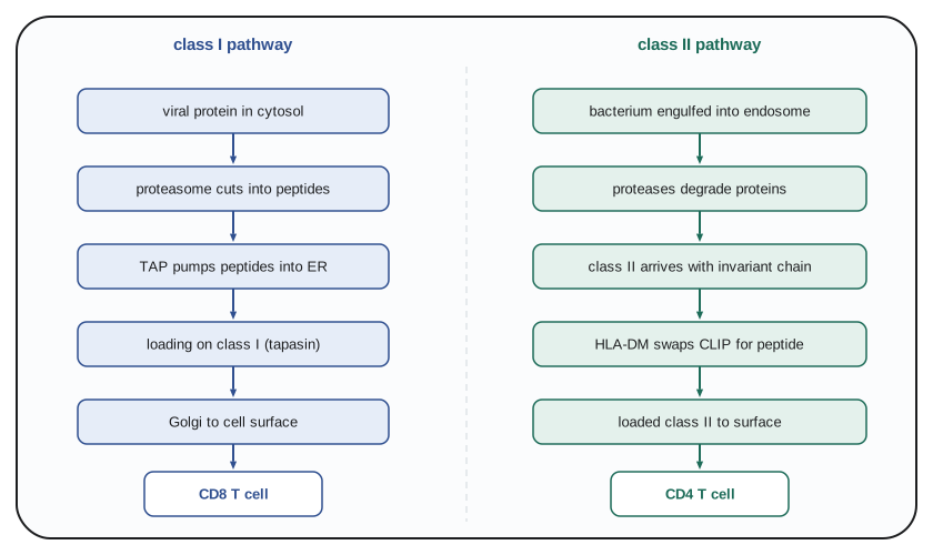
Every cell constantly degrades its own proteins. In the class I pathway, cytosolic and nuclear proteins, including viral proteins in an infected cell and mutant proteins in a cancer cell, are tagged with ubiquitin and broken down by the proteasome, a barrel-shaped protease complex. Interferon-γ induces replacement of three proteasome subunits to form the immunoproteasome, which produces more peptides with the hydrophobic or basic carboxyl ends that class I molecules prefer. The peptides are pumped into the endoplasmic reticulum by the transporter associated with antigen processing (TAP), a heterodimer of TAP1 and TAP2. Inside the ER, newly made class I molecules are held in a peptide-loading complex with chaperones, including calreticulin, ERp57 and tapasin, which bridges class I to TAP and helps select peptides that bind stably. ER aminopeptidases trim peptides to the right length. Once a peptide binds, the class I molecule is released and travels through the Golgi to the cell surface.
A healthy cell's class I molecules display a sample of thousands of different self peptides, each in a few to a few thousand copies; a virus-infected cell adds viral peptides to the mix. Most defective ribosomal products, proteins that misfold or terminate early during synthesis, are degraded almost immediately, which means that viral peptides can appear on the surface within an hour or two of infection, long before new virus particles are released. The importance of TAP is shown by people with rare TAP deficiency, who have very low surface class I and suffer chronic bacterial infections of the airways and skin ulcers, and by the many viruses that block TAP: herpes simplex virus makes a protein, ICP47, that plugs it, and human cytomegalovirus makes several proteins that destroy class I molecules or retain them in the ER. Such evasion makes infected cells vulnerable to NK cells, which detect missing self.
The class II pathway: presenting what the cell has engulfed
In the class II pathway, antigen-presenting cells take up extracellular material by phagocytosis, macropinocytosis or receptor-mediated endocytosis; B cells take up antigen bound to their antibody receptors with great efficiency. The material is degraded by proteases such as cathepsins in increasingly acidic endosomes. Newly made class II molecules assemble in the ER with a third protein, the invariant chain (CD74), part of which occupies the peptide groove and prevents ER peptides from binding. The invariant chain also carries a signal that directs class II to late endosomes. There, proteases degrade the invariant chain, leaving a fragment called CLIP in the groove. The chaperone HLA-DM catalyzes the exchange of CLIP for antigenic peptides, favoring those that bind tightly; a related molecule, HLA-DO, modulates DM in B cells. Loaded class II molecules then travel to the cell surface.
Autophagy, introduced in BIOL-214, links the two pathways: it delivers cytosolic proteins to lysosomes, where they can be loaded onto class II molecules. This route allows CD4 T cells to recognize some viral and self proteins that are made inside the cell, and it is important for presenting self antigens in the thymus during T cell development.
Cross-presentation
The division of labor between the two pathways creates a puzzle. To start a CD8 T cell response against a virus, a dendritic cell must display viral peptides on class I. But many viruses do not infect dendritic cells, and those that do often disable them. How, then, are CD8 T cells primed? The answer is cross-presentation: certain dendritic cells can take up material from other cells, such as dying virus-infected or tumor cells, and route it into the class I pathway. Michael Bevan described the phenomenon in 1976. In mice and humans a particular subset, conventional type 1 dendritic cells (cDC1), marked by the receptor XCR1 and, in mice, CD8α, is specialized for cross-presentation, using mechanisms that include transport of engulfed proteins from endosomes into the cytosol for proteasomal degradation. Cross-presentation is essential for CD8 T cell responses to many viruses and to tumors, and mice lacking cDC1 cells cannot reject transplanted tumors, which has made these cells a focus of cancer immunotherapy research.
Dendritic cells: the gatekeepers
Dendritic cells are uniquely able to activate naive T cells. In tissues, immature dendritic cells are highly endocytic, continually sampling their surroundings, but they express modest levels of MHC and costimulatory molecules. When their pattern recognition receptors detect microbial products or tissue damage, they mature: they stop taking up antigen, increase surface MHC class II and the costimulatory molecules CD80 and CD86, switch their chemokine receptors to CCR7, which guides them through lymphatics to the T cell zones of lymph nodes, and produce cytokines such as interleukin-12. A mature dendritic cell arriving in a lymph node therefore displays peptides from the place it came from, together with signals that report danger, and presents them to the many naive T cells that pass through each day.
The requirement for this maturation is a safeguard. Dendritic cells that present self antigens without maturation signals, as they do continually in the steady state, tend to make T cells unresponsive or convert them to regulatory T cells rather than activating them, which helps maintain tolerance (lesson 8). It also explains why vaccines need adjuvants: purified proteins alone rarely provoke strong responses, because they do not trigger dendritic cell maturation. Several dendritic cell types exist, including cDC1 cells for cross-presentation and CD8 responses, cDC2 cells, which are efficient at activating CD4 T cells, and plasmacytoid dendritic cells, which make large amounts of type I interferon, together with Langerhans cells in the epidermis, now thought to be closer to macrophages in origin.
The human HLA complex and its diversity
The human MHC, on the short arm of chromosome 6, spans about four million base pairs and contains more than 200 genes, many with immune functions. The classical class I genes are HLA-A, HLA-B and HLA-C; the classical class II genes encode the DR, DQ and DP molecules. Between them lie genes for complement components, TNF and other immune proteins, sometimes called the class III region. Because the genes are close together, they are usually inherited as a block, called a haplotype, one from each parent.
The HLA genes are the most polymorphic in the human genome. The IPD-IMGT/HLA database, the international reference, lists more than 40,000 HLA alleles, with HLA-B alone having more than 10,000. Most of the variation lies in the residues lining the peptide groove, so different alleles bind different sets of peptides. Because HLA genes are codominantly expressed, a person typically has up to six different classical class I molecules (two each of A, B and C) and several different class II molecules, including those formed by pairing of α and β chains from different parents in the case of DQ and DP.
Why is HLA so diverse? The prevailing explanation is that pathogens drive balancing selection. A person heterozygous at HLA loci can present a wider range of peptides from any pathogen, which should improve resistance; this heterozygote advantage has been observed in HIV infection, where people heterozygous at all class I loci progress to AIDS more slowly. In addition, rare alleles may be favored because pathogens adapt to evade presentation by common ones, a form of negative frequency-dependent selection. At the population level, diversity reduces the chance that a single pathogen can escape presentation in everyone. Some HLA lineages are older than the human species, shared with chimpanzees, and modern humans acquired some alleles by interbreeding with Neanderthals and Denisovans, as discussed in BIOL-322.
import random
# HLA matching between siblings versus unrelated people (illustrative simulation).
# Each parent carries two haplotypes; a child inherits one at random from each parent.
random.seed(380)
def sib_match(trials=100000):
    hits = 0
    for _ in range(trials):
        a = (random.randint(0, 1), random.randint(0, 1))
        b = (random.randint(0, 1), random.randint(0, 1))
        hits += a == b
    return hits / trials
print(f"chance two siblings share both haplotypes: {sib_match():.3f} (expected 0.25)")
n_sibs = [1, 2, 3, 4, 5]
for n in n_sibs:
    print(f"  patient with {n} sibling(s): chance at least one is a full match = {1 - 0.75 ** n:.2f}")
# Unrelated donors: illustrative haplotype frequencies (real ones vary greatly by ancestry).
freqs = {"common haplotype (5%)": 0.05, "moderately common (1%)": 0.01,
         "uncommon (0.1%)": 0.001, "rare (0.01%)": 0.0001}
registry = 40_000_000   # order of magnitude of adult donors in worldwide registries
print("\nchance that a random unrelated donor carries both of the patient's haplotypes:")
for name, f in freqs.items():
    p = 2 * f * f      # two different haplotypes, each with frequency f: genotype frequency 2f^2
    expected = p * registry
print(f" {name:24s} per-donor probability {p:.1e}, expected matches in registry {expected:,.1f}")chance two siblings share both haplotypes: 0.249 (expected 0.25) patient with 1 sibling(s): chance at least one is a full match = 0.25 patient with 2 sibling(s): chance at least one is a full match = 0.44 patient with 3 sibling(s): chance at least one is a full match = 0.58 patient with 4 sibling(s): chance at least one is a full match = 0.68 patient with 5 sibling(s): chance at least one is a full match = 0.76 chance that a random unrelated donor carries both of the patient's haplotypes: common haplotype (5%) per-donor probability 5.0e-03, expected matches in registry 200,000.0 moderately common (1%) per-donor probability 2.0e-04, expected matches in registry 8,000.0 uncommon (0.1%) per-donor probability 2.0e-06, expected matches in registry 80.0 rare (0.01%) per-donor probability 2.0e-08, expected matches in registry 0.8
The simulation shows the arithmetic behind transplant matching. A sibling inherits the same pair of parental haplotypes with probability one in four, so a patient with two siblings has a 44 percent chance that at least one is a full match. For an unrelated donor, the chance of a match depends on how common the patient's haplotypes are: common haplotypes may be matched many times over in registries of tens of millions, while rare ones may have no match at all. The calculation above simplifies by assuming both of the patient's haplotypes have the same frequency, that genotypes follow Hardy–Weinberg proportions, and by ignoring partial matches, which are often acceptable. Because haplotype frequencies differ by ancestry and registries are dominated by donors of European descent, patients of African, Indigenous, Asian and mixed ancestry are less likely to find a fully matched unrelated donor, one reason registries such as Canadian Blood Services' Stem Cell Registry and the US National Marrow Donor Program actively recruit diverse donors.
HLA and disease
Particular HLA alleles are associated with susceptibility to many diseases, especially autoimmune diseases. The strongest association known is between HLA-B27 and ankylosing spondylitis, an inflammatory disease of the spine: more than 90 percent of patients of European descent carry B27, compared with about 8 percent of the general population. Celiac disease almost always occurs in people carrying HLA-DQ2 or DQ8, which bind gluten peptides that have been modified by the enzyme tissue transglutaminase. Type 1 diabetes is associated with DR3 and DR4 haplotypes carrying particular DQ alleles, and multiple sclerosis with HLA-DRB1*15:01. HLA alleles also predict severe reactions to certain drugs: carriers of HLA-B*57:01 are at high risk of a hypersensitivity reaction to the HIV drug abacavir, so patients are tested before treatment, and HLA-B*15:02, common in parts of Southeast Asia, predicts severe skin reactions to the anticonvulsant carbamazepine. These are among the clearest examples of pharmacogenomics in routine use, covered further in PHAR-361.
Transplantation antigens
The polymorphism of HLA is the main barrier to transplantation. Between 1 and 10 percent of a person's T cells respond to any single foreign HLA molecule, far more than the one in 10^5 or so that responds to a given foreign peptide on self MHC. This high frequency of alloreactive T cells arises because each foreign MHC molecule carrying many different peptides resembles a large number of self-MHC–peptide combinations that T cells were selected to recognize. Lesson 11 examines how this leads to rejection and how matching and immunosuppression prevent it.
Worked problem
Predicting which T cells respond
A cell line infected with influenza virus is treated with a drug that blocks TAP. A second sample is treated with chloroquine, which raises endosomal pH and blocks endosomal proteases. Which T cell responses to influenza peptides will each treatment impair, and why?
Show solution
- Influenza proteins are made in the cytosol of the infected cell, so their peptides normally reach class I through the proteasome and TAP.
- Blocking TAP prevents cytosolic peptides from entering the ER, so class I molecules cannot load viral peptides: CD8 T cell recognition is impaired.
- Chloroquine blocks endosomal processing, which is required for loading class II: CD4 T cell recognition of engulfed influenza antigen is impaired, while class I presentation of newly synthesized viral proteins continues.
- Some cross-talk exists, such as autophagy delivering cytosolic proteins to class II and cross-presentation in dendritic cells, so the effects in real experiments are rarely absolute.
Answer: TAP blockade impairs CD8 T cell recognition through class I; chloroquine impairs CD4 T cell recognition through class II.
Common mistakes
MistakeThinking T cells recognize whole native proteins.
InsteadT cell receptors recognize short peptides bound to MHC molecules.
MistakePlacing MHC class II on all nucleated cells.
InsteadClass II is mainly on professional antigen-presenting cells; class I is on almost all nucleated cells.
MistakeAssuming CD8 T cells can only be primed by infected dendritic cells.
InsteadCross-presentation lets dendritic cells load class I with antigens taken up from other cells.
MistakeBelieving each person has one HLA molecule per class.
InsteadCodominant expression gives up to six classical class I molecules and several class II molecules.
MistakeDescribing HLA polymorphism as random noise.
InsteadIt is maintained by pathogen-driven balancing selection and concentrated in the peptide-binding groove.
Summary and key terms
- T cells recognize peptides bound to self MHC molecules (MHC restriction), discovered by Zinkernagel and Doherty.
- Class I molecules on almost all nucleated cells present cytosolic peptides of 8–10 residues to CD8 T cells.
- Class II molecules on antigen-presenting cells present endosomal peptides of 13–25 residues to CD4 T cells.
- The class I pathway uses the proteasome, TAP and the peptide-loading complex; the class II pathway uses invariant chain, CLIP and HLA-DM.
- Cross-presentation by cDC1 dendritic cells lets CD8 T cells respond to antigens from other cells.
- Dendritic cells mature on sensing danger and migrate to lymph nodes to activate naive T cells.
- HLA genes are extraordinarily polymorphic, shaping disease susceptibility, drug reactions and transplant matching.
- major histocompatibility complex
- A gene region encoding molecules that present peptides to T cells.
- HLA
- Human leukocyte antigen, the human MHC.
- MHC restriction
- The requirement that T cells recognize antigen bound to self MHC.
- anchor residue
- A peptide side chain that fits a specific pocket in the MHC groove.
- proteasome
- A cytosolic protease complex that degrades ubiquitinated proteins into peptides.
- TAP
- Transporter associated with antigen processing, which pumps peptides into the ER.
- invariant chain
- A protein that blocks the class II groove and directs class II to endosomes.
- HLA-DM
- A chaperone that exchanges CLIP for antigenic peptides on class II.
- cross-presentation
- Presentation of exogenous antigens on MHC class I by dendritic cells.
- haplotype
- A set of linked alleles inherited together on one chromosome.
- alloreactive T cell
- A T cell that responds to foreign MHC molecules from another individual.
Practice problems
Work each problem on paper first, then reveal the answer to check.
What did Zinkernagel and Doherty discover?
Show answer
That cytotoxic T cells recognize viral antigen only together with self MHC.
Why does the class I groove bind shorter peptides than class II?
Show answer
It is closed at both ends, so the peptide termini must fit within it.
Name the steps that bring a viral peptide to class I.
Show answer
Ubiquitination, proteasomal degradation, TAP transport into the ER, loading by the peptide-loading complex, export to the surface.
What is the role of the invariant chain?
Show answer
It blocks the class II groove in the ER and targets class II to endosomes.
Why is cross-presentation needed?
Show answer
To prime CD8 T cells against viruses that do not infect dendritic cells, and against tumors.
What is the chance that one sibling is a full HLA match, and why?
Show answer
One in four, because each parent passes one of two haplotypes.
Give two examples of HLA alleles that predict drug reactions.
Show answer
HLA-B*57:01 and abacavir; HLA-B*15:02 and carbamazepine.
Why are alloreactive T cells so common?
Show answer
Each foreign MHC molecule with its many peptides resembles many self-MHC–peptide complexes T cells were selected on.
Lesson quiz · 5 questions
Score 60% or more to complete this lesson.
Create a free account to save your progress, take quizzes and earn a certificate.