
BIOL-380 · Lesson 1 of 12 · 30 min
The immune system: cells, organs and principles
Every animal lives surrounded by microbes, and many of them would happily grow in its tissues. The immune system is the collection of cells, molecules and organs that prevents this, and it does so while sparing the body's own tissues and the trillions of harmless microbes that live on the skin and in the gut. This course examines how that system works at the level a third-year biologist needs: how it recognizes what is dangerous, how it builds a response suited to each threat, how it remembers past encounters, and how it is kept in check. This first lesson sets out the main principles, the cells involved and the organs where they develop and meet. Later lessons take each part in turn, then examine what happens when immunity fails or turns against the body, and how vaccines and immunotherapies harness it.
The course builds on cell biology from BIOL-214, which covered receptors, signaling pathways, the cell cycle and membrane traffic, and on molecular biology from BIOL-207. Microbiology from BIOL-330 introduced the pathogens themselves, including bacterial cell walls, viral replication and antimicrobial resistance; here the focus is on the host. Clinical detail on drugs used to treat immune disorders belongs to the pharmacy and medicine courses, and this course points to them where relevant.
A short history of immunology
People noticed long before anyone understood why that survivors of some diseases did not catch them again. In fifth-century BCE Athens, the historian Thucydides recorded that those who had recovered from the plague could nurse the sick without falling ill a second time. Variolation, the deliberate infection of healthy people with material from smallpox sores to produce a milder disease and lasting protection, was practiced in China, India and the Ottoman Empire for centuries and was brought to Britain in the 1720s by Lady Mary Wortley Montagu. In 1796 Edward Jenner showed that inoculation with material from cowpox lesions protected against smallpox at far lower risk; the word vaccine comes from the Latin vacca, cow.
Louis Pasteur extended the idea in the 1880s, producing attenuated vaccines against chicken cholera, anthrax and rabies, and generalized Jenner's term to honor him. At about the same time two schools argued over how immunity worked. Élie Metchnikoff, watching mobile cells in starfish larvae engulf thorns, proposed that phagocytic cells were the main defense. Emil von Behring and Shibasaburo Kitasato showed in 1890 that serum from animals immunized against diphtheria or tetanus toxin could protect other animals, implying that soluble substances, later called antibodies, did the work. Paul Ehrlich proposed a side-chain theory in which cells carried receptors that were shed as antibodies. Both camps were right: Metchnikoff and Ehrlich shared the Nobel Prize in 1908, and modern immunology describes the cooperation of cells and soluble molecules.
The central idea of adaptive immunity came from Frank Macfarlane Burnet in 1957. His clonal selection theory proposed that each lymphocyte carries receptors of a single specificity, generated before any antigen is met; that antigen selects the rare cells whose receptors bind it and causes them to divide into a clone; and that lymphocytes that react with self are eliminated during development. Every one of these predictions has been confirmed, and the theory remains the framework of the field. Burnet also framed immunity as discrimination between self and non-self. In 1989 Charles Janeway pointed out that the adaptive system usually responds only when the innate system signals the presence of microbes, and in 1994 Polly Matzinger proposed the danger model, in which the immune system responds to signs of tissue damage and stress rather than to foreignness as such. These ideas are not mutually exclusive, and each explains observations the others struggle with, such as tolerance of the fetus, of food and of the gut microbiota.
Innate and adaptive immunity
Immunologists divide the immune system into two interlocking arms. Innate immunity is present before infection, responds within minutes to hours, and recognizes broad classes of molecules shared by many microbes, such as bacterial cell wall components, flagellin and double-stranded viral RNA, through receptors encoded in the germline. It includes physical and chemical barriers, the complement system of plasma proteins, and cells such as neutrophils, macrophages, dendritic cells and natural killer cells. Innate responses do not, in the classical view, improve with repeated exposure, although lesson 3 describes evidence for a form of innate memory called trained immunity.
Adaptive immunity is carried out by lymphocytes, the B cells and T cells, whose antigen receptors are assembled by gene rearrangement during development, so that each cell has a different receptor and the population as a whole can recognize almost any molecular shape. An adaptive response takes days to develop on first exposure, because the few cells with suitable receptors must be activated and must proliferate; but it is highly specific, and it leaves behind memory cells that respond faster and more strongly to a second exposure. This combination of specificity and memory is what vaccines exploit.
| Feature | Innate immunity | Adaptive immunity |
|---|---|---|
| Timing of first response | Minutes to hours | Days (about 4 to 7 for a primary response) |
| Receptors | Germline-encoded pattern recognition receptors, the same on all cells of a type | Rearranged B and T cell receptors, unique to each clone |
| What is recognized | Conserved microbial and damage-associated molecular patterns | Specific epitopes on almost any molecule (antibodies) or peptides on MHC (T cells) |
| Diversity of receptors | Hundreds of receptor types | Potentially more than 10^13 distinct receptors |
| Memory | Limited (trained immunity) | Strong, long-lived memory cells |
| Main cells | Neutrophils, macrophages, dendritic cells, mast cells, NK cells, innate lymphoid cells | B cells, CD4 and CD8 T cells |
| Main soluble molecules | Complement, cytokines, antimicrobial peptides, interferons | Antibodies, cytokines |
The two arms are not separate systems. Innate cells decide whether an adaptive response starts at all and what kind it will be: dendritic cells carry antigen to lymph nodes and display it to T cells together with signals that report what sort of microbe was found. Adaptive responses, in turn, focus and amplify innate mechanisms. Antibodies coat microbes so that phagocytes can engulf them and so that complement is activated on their surface; T cells release cytokines that make macrophages better killers. Much of immunology is the study of this conversation.
The cells of the immune system
All blood cells, including immune cells, descend from hematopoietic stem cells in the bone marrow. These rare, self-renewing cells give rise to two main lineages. The common myeloid progenitor produces red blood cells, platelets, granulocytes, monocytes and most dendritic cells; the common lymphoid progenitor produces B cells, T cells, natural killer cells and the innate lymphoid cells. Hematopoiesis is controlled by cytokines and growth factors such as granulocyte colony-stimulating factor, which is given to patients after chemotherapy to speed the recovery of neutrophils. An adult produces roughly 10^11 neutrophils a day, a striking figure that reflects how short-lived they are.
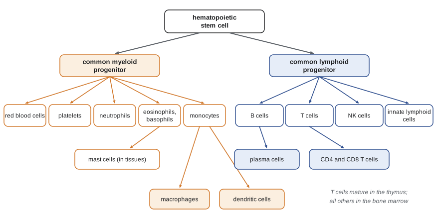
Granulocytes are named for the granules in their cytoplasm. Neutrophils are the most abundant white blood cells, making up about half to two thirds of leukocytes in the blood, and they are the first cells to arrive at a site of bacterial infection, where they engulf and kill microbes and then die within a day or two; pus consists largely of dead neutrophils. Eosinophils defend against parasitic worms and contribute to allergic inflammation, and basophils and the tissue-resident mast cells release histamine and other mediators that cause the immediate symptoms of allergy, examined in lesson 10.
Monocytes circulate in the blood and enter tissues, where they mature into macrophages. Many tissues also contain resident macrophages that were seeded during embryonic development and renew themselves locally, such as the Kupffer cells of the liver, the alveolar macrophages of the lung and the microglia of the brain. Macrophages engulf microbes and dead cells, release cytokines that orchestrate inflammation and later help repair tissue. Dendritic cells, named for their long branching processes, sample antigens in tissues, migrate to lymph nodes and present antigen to naive T cells; they are the essential link between innate and adaptive immunity, a discovery for which Ralph Steinman of Rockefeller University, a graduate of McGill University, received the Nobel Prize in 2011, awarded three days after his death.
Lymphocytes look alike under the light microscope, small round cells with a large nucleus and little cytoplasm, but they differ greatly in function. B cells mature in the bone marrow and, when activated, become plasma cells that secrete antibodies. T cells mature in the thymus and come in two main types: CD4 T cells, also called helper T cells, which coordinate responses through cytokines and direct contact, and CD8 T cells, also called cytotoxic T lymphocytes, which kill infected and cancerous cells. Natural killer cells are innate lymphocytes that kill stressed and virus-infected cells without needing prior sensitization, and the innate lymphoid cells, discovered only in the past fifteen years, are tissue-resident cells that mirror the cytokine profiles of helper T cells but lack rearranged receptors.
Immune cells are identified by the molecules on their surface. Since the 1980s these markers have been given cluster of differentiation (CD) numbers, assigned at international workshops, and more than 370 are now named. CD4 and CD8 mark the two main T cell classes, CD19 and CD20 mark B cells, CD3 is part of the T cell receptor complex, CD56 marks NK cells, and CD45 is found on all leukocytes. Lesson 11 shows how flow cytometry uses fluorescent antibodies against these markers to count cell types, which is how the CD4 count of a person with HIV is measured.
The organs of the immune system
The primary lymphoid organs are where lymphocytes develop. The bone marrow produces all blood cells and is where B cells mature; the thymus, a two-lobed organ above the heart, is where T cells mature. The thymus is largest relative to body size in infancy and shrinks after puberty, a process called involution, so that the output of new T cells falls with age, although it never stops entirely. Lesson 6 describes the selection that takes place there, which removes most developing T cells.
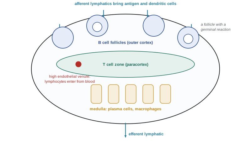
The secondary lymphoid organs are where mature lymphocytes meet antigen and are activated. Lymph nodes, roughly 450 to 600 of them in an adult human, sit along lymphatic vessels that drain fluid from the tissues. Lymph carries antigens and migrating dendritic cells from the tissues into the node, while naive lymphocytes enter from the blood through specialized high endothelial venules. Inside the node, B cells cluster in follicles in the outer cortex and T cells in the paracortex beneath, and each naive lymphocyte spends several hours in a node scanning for its antigen before leaving through the efferent lymph to try another. The spleen filters the blood in the same way, and also removes old red blood cells, which is why people without a spleen are at particular risk from encapsulated bacteria such as Streptococcus pneumoniae carried in the bloodstream.
Mucosal surfaces, which in a human adult cover an area of roughly 30 to 40 square meters (older estimates of several hundred square meters counted every microvillus), are protected by mucosa-associated lymphoid tissue (MALT). This includes the tonsils and adenoids, the Peyer's patches of the small intestine and scattered lymphoid follicles in the airways and gut. Specialized M cells in the epithelium above Peyer's patches transport antigens from the gut lumen to underlying immune cells. The mucosal immune system must tolerate food and commensal microbes while responding to pathogens, and its antibody of choice is IgA, secreted across the epithelium in enormous quantities, several grams a day in an adult.
Principles of immune recognition
Five principles recur throughout the course. The first is specificity: each adaptive receptor binds a particular molecular shape, the epitope or antigenic determinant, and a typical protein carries many epitopes, so the response to a microbe involves many clones. The second is diversity: the repertoire of B and T cell receptors is generated randomly before antigen is met, so it must be large enough that some cells will recognize almost anything. The third is clonal expansion: because only about one naive T cell in 10^5 to 10^6 recognizes a given epitope, the response depends on rapid proliferation of those few cells, which can divide every six to eight hours once activated.
The fourth principle is memory: after an infection is cleared, most of the expanded clone dies, but some cells persist as memory cells that are more numerous and more easily activated than the original naive cells. The fifth is regulation and tolerance: the receptors that are generated randomly will inevitably include some that recognize self, so the system has mechanisms to delete or silence these cells and to shut responses down once the threat has passed. Failure of tolerance causes autoimmune disease, and excessive or misdirected responses cause allergy and other forms of hypersensitivity.
import math
# Clonal expansion: how long does it take a rare naive clone to become a large effector population?
# Illustrative values: precursor frequency, number of naive CD8 T cells, division time.
naive_cd8 = 1.0e11          # naive CD8 T cells in an adult (order of magnitude)
for freq in (1e-5, 1e-6):
    precursors = naive_cd8 * freq
    print(f"precursor frequency 1 in {1 / freq:,.0f}: about {precursors:,.0f} naive cells specific for the epitope")
target = 1.0e10             # effector cells at the peak of a strong response (order of magnitude)
for div_hours in (6, 8, 12):
    precursors = naive_cd8 * 1e-6
    n_div = math.log2(target / precursors)
print(f"division every {div_hours} h: {n_div:.1f} divisions = {n_div * div_hours / 24:.1f} days after a lag to reach {target:.0e} cells")precursor frequency 1 in 100,000: about 1,000,000 naive cells specific for the epitope precursor frequency 1 in 1,000,000: about 100,000 naive cells specific for the epitope division every 6 h: 16.6 divisions = 4.2 days after a lag to reach 1e+10 cells division every 8 h: 16.6 divisions = 5.5 days after a lag to reach 1e+10 cells division every 12 h: 16.6 divisions = 8.3 days after a lag to reach 1e+10 cells
The calculation shows why a primary response peaks about a week after infection. Starting from about 100,000 cells specific for an epitope, roughly 17 divisions are needed to reach ten billion, and at six to eight hours per division that takes four to six days, after an initial lag of a day or two while dendritic cells migrate and T cells are activated. Real responses vary widely, but measurements in mice and humans after acute viral infections and vaccines, such as the yellow fever vaccine, show expansions of this order, with a single epitope-specific population sometimes making up a tenth or more of all CD8 T cells at the peak.
The phases of an immune response
A typical response to a bacterial skin infection illustrates how the parts fit together. Bacteria entering through a cut first meet barriers: the skin, antimicrobial peptides and complement. Those that survive are recognized within minutes by resident macrophages and mast cells, which release cytokines and other mediators that dilate blood vessels and attract neutrophils from the blood, producing the redness, heat, swelling and pain of inflammation. Complement proteins coat the bacteria, making them easier to engulf, and some complement fragments punch holes in bacterial membranes.
Meanwhile, dendritic cells in the skin take up bacterial antigens, become activated, and migrate through lymphatics to the draining lymph node, arriving within a day or two. There they present bacterial peptides on MHC molecules to naive T cells; the rare cells whose receptors fit are activated, proliferate and differentiate. Helper T cells move to the edge of the B cell follicles and help B cells that have bound the same antigen to proliferate, switch antibody class and improve their affinity. Over the following days, antibodies and effector T cells leave the node and travel to the site of infection, where they help clear the bacteria. Once the infection is controlled, most effector cells die, inflammation resolves, tissue is repaired, and memory cells remain. If the same bacterium returns, the memory response is faster, larger and of higher affinity, often clearing the infection before symptoms appear.
Immunology in Canada and the United States
American scientists have made many landmark contributions to immunology. Mark Davis's group at Stanford cloned the mouse T cell receptor beta chain in 1984, at the same time as Tak Mak's group in Toronto cloned the human version; Ralph Steinman discovered dendritic cells at Rockefeller University; and James Allison, working at Berkeley and later MD Anderson Cancer Center, developed checkpoint blockade, described in lesson 12. Research is funded largely by the National Institutes of Health, in particular the National Institute of Allergy and Infectious Diseases. Vaccine recommendations have been made by the Advisory Committee on Immunization Practices (ACIP), which advises the Centers for Disease Control and Prevention; lesson 9 describes the substantial changes to that process since 2025.
Worked problem
Interpreting a white blood cell count
A routine blood test reports a total white blood cell count of 12.0 × 10^9 cells per liter, with 80 percent neutrophils, 12 percent lymphocytes, 6 percent monocytes and 2 percent eosinophils. A typical adult reference range is about 4 to 11 × 10^9 per liter total, with neutrophils about 2 to 7.5 × 10^9 per liter. What do these figures suggest?
Show solution
- Absolute neutrophil count = 0.80 × 12.0 = 9.6 × 10^9 per liter, above the reference range (neutrophilia).
- Absolute lymphocyte count = 0.12 × 12.0 = 1.44 × 10^9 per liter, within or near the low end of normal.
- A raised total count driven by neutrophils is typical of an acute bacterial infection or other acute inflammation, because the bone marrow releases stored neutrophils and increases production.
- Absolute counts, not percentages, should be interpreted: a high percentage of neutrophils can coexist with a normal absolute count if the total is low.
Answer: The patient has neutrophilia, consistent with an acute bacterial infection or other inflammatory process; clinicians interpret such results together with symptoms and other tests.
Common mistakes
MistakeThinking innate immunity is crude and adaptive immunity sophisticated.
InsteadInnate immunity uses precise receptors for conserved microbial molecules and decides whether and how adaptive responses start.
MistakeAssuming each lymphocyte can recognize many antigens.
InsteadEach B or T cell carries receptors of a single specificity; diversity comes from the population.
MistakeBelieving the receptor repertoire is created in response to antigen.
InsteadReceptors are generated randomly before antigen is met; antigen selects existing clones (clonal selection).
MistakeTreating the thymus and bone marrow as sites of immune responses.
InsteadThey are primary organs where lymphocytes develop; responses start in secondary organs such as lymph nodes and spleen.
MistakeReading white cell percentages without converting to absolute counts.
InsteadMultiply each percentage by the total count before comparing with reference ranges.
Summary and key terms
- Immunology grew from observations of lasting protection after illness, through variolation, Jenner and Pasteur, to Burnet's clonal selection theory.
- Innate immunity is rapid and uses germline-encoded receptors; adaptive immunity is slower, highly specific and has memory.
- All immune cells derive from hematopoietic stem cells through myeloid and lymphoid lineages.
- Primary lymphoid organs (bone marrow, thymus) produce lymphocytes; secondary organs (lymph nodes, spleen, MALT) are where responses begin.
- Specificity, diversity, clonal expansion, memory and tolerance are the core principles of adaptive immunity.
- A response proceeds from barriers and innate recognition to antigen presentation in lymph nodes, clonal expansion, effector function, contraction and memory.
- antigen
- Any molecule recognized by an antibody or T cell receptor.
- epitope
- The part of an antigen bound by a single antibody or T cell receptor.
- innate immunity
- Defenses present before infection that recognize conserved microbial molecules through germline-encoded receptors.
- adaptive immunity
- Responses by B and T cells with rearranged receptors, showing specificity and memory.
- clonal selection
- The selection and expansion of pre-existing lymphocytes whose receptors bind an antigen.
- hematopoietic stem cell
- A self-renewing bone marrow cell that gives rise to all blood cells.
- dendritic cell
- An antigen-presenting cell that carries antigen to lymph nodes and activates naive T cells.
- primary lymphoid organ
- An organ where lymphocytes develop: the bone marrow and thymus.
- secondary lymphoid organ
- An organ where lymphocytes meet antigen and are activated, such as lymph nodes and the spleen.
- cluster of differentiation
- A numbered system for naming cell-surface molecules used to identify cell types.
Practice problems
Work each problem on paper first, then reveal the answer to check.
What did Burnet's clonal selection theory predict?
Show answer
Each lymphocyte has one specificity generated before antigen exposure; antigen selects and expands matching clones; self-reactive cells are removed.
Name three differences between innate and adaptive immunity.
Show answer
Speed, receptor type (germline versus rearranged) and memory.
Which cells link innate and adaptive immunity, and how?
Show answer
Dendritic cells carry antigen to lymph nodes and present it to naive T cells with activating signals.
Where do B cells and T cells mature?
Show answer
B cells in the bone marrow, T cells in the thymus.
Why are people without a spleen vulnerable to encapsulated bacteria in the blood?
Show answer
The spleen filters blood and mounts responses to blood-borne bacteria.
About how many divisions turn 100,000 cells into 10 billion?
Show answer
About 17, since 2^17 is roughly 130,000.
What antibody class dominates at mucosal surfaces?
Show answer
IgA.
Calculate the absolute lymphocyte count when the total is 6 × 10^9 per liter and lymphocytes are 30 percent.
Show answer
1.8 × 10^9 per liter.
Lesson quiz · 5 questions
Score 60% or more to complete this lesson.
Create a free account to save your progress, take quizzes and earn a certificate.