
BIOL-380 · Lesson 2 of 12 · 28 min
Innate immunity I: barriers, complement and recognition
Most microbes that touch the body never cause an infection, because they are stopped by defenses that are always in place. This lesson examines the first layers of protection: the physical, chemical and microbial barriers of the skin and mucosa; the complement system, a cascade of plasma proteins that marks and destroys microbes within minutes; and the receptors with which innate cells recognize that something foreign or dangerous is present. Lesson 3 then follows what happens once recognition has occurred: inflammation, cytokines and the work of innate cells.
Barriers
The skin is a remarkably effective barrier. Its outer layer, the stratum corneum, consists of flattened dead keratinocytes embedded in lipids, a dry, acidic and constantly shedding surface on which few pathogens can gain a foothold. Sebaceous glands secrete fatty acids that inhibit bacteria, and sweat contains salt, lysozyme and the antimicrobial peptide dermcidin. Breaks in the skin, from cuts, burns, insect bites or medical devices such as catheters, are the main routes by which skin bacteria cause infection, which is why burns patients are so vulnerable.
Mucosal surfaces of the gut, airways and urogenital tract are far larger and thinner, often a single layer of epithelial cells, and they must allow exchange of gases, nutrients and fluids. Their defenses are correspondingly active. Goblet cells secrete mucus, a gel of heavily glycosylated mucin proteins that traps microbes and keeps them away from the epithelium; in the colon an inner mucus layer is almost sterile. In the airways, cilia beat the mucus upward at a few millimeters per minute toward the throat, where it is swallowed, the mucociliary escalator that smoking and the genetic disease cystic fibrosis impair. Tears, saliva and urine flush surfaces, the stomach's acidity kills many ingested microbes, and tight junctions seal the gaps between epithelial cells.
Epithelial cells and neutrophils also produce antimicrobial peptides, small cationic molecules of roughly 12 to 50 amino acids that bind to the negatively charged membranes of bacteria, fungi and some enveloped viruses and disrupt them. Humans make two main families: the defensins, including alpha-defensins secreted by Paneth cells at the base of intestinal crypts, and the cathelicidin LL-37. Because they attack membranes broadly, resistance to them evolves slowly, which has made them candidates for new antibiotics. Other antimicrobial proteins include lysozyme, which cleaves the peptidoglycan of bacterial cell walls, lactoferrin, which sequesters iron, and the C-type lectin RegIIIγ in the gut, which kills Gram-positive bacteria.
The microbiota is itself a barrier. The trillions of bacteria that live in the gut and on the skin compete with pathogens for nutrients and attachment sites, produce bacteriocins and short-chain fatty acids that inhibit them, and train the immune system. When antibiotics disrupt the gut microbiota, the spore-forming bacterium Clostridioides difficile can expand and cause severe colitis; transplantation of fecal microbiota from healthy donors cures most recurrent cases, a striking demonstration of colonization resistance. BIOL-330 covered the microbiome in more detail.
The complement system
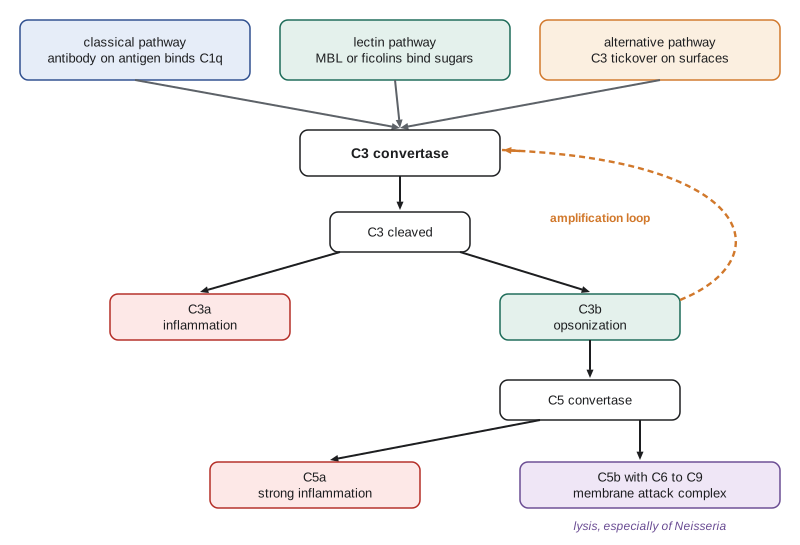
Complement was discovered in the 1890s by Jules Bordet, who showed that a heat-sensitive component of fresh serum complemented the action of antibodies in killing bacteria. It is now known to comprise more than 30 plasma and membrane proteins, most made by the liver, that circulate in inactive forms. When activated, many of them become proteases that cleave and activate the next component, so that a small trigger is amplified into a large response, a principle similar to blood clotting. Complement has three main effects: opsonization, in which fragments of the protein C3 coat a microbe and target it for phagocytosis; inflammation, in which small fragments called anaphylatoxins attract and activate leukocytes; and lysis, in which a membrane attack complex punches holes in the target's membrane.
Three pathways converge on the cleavage of C3, the most abundant complement protein, present in plasma at about 1 to 1.5 grams per liter. The classical pathway, the first discovered, begins when the protein C1q binds to antibodies, IgM or certain IgG subclasses, attached to a microbe; it links complement to adaptive immunity. The lectin pathway begins when mannose-binding lectin or ficolins bind carbohydrate patterns on microbial surfaces, such as the mannose-rich glycans of yeasts and many bacteria. Both pathways then cleave the proteins C4 and C2 to form the classical C3 convertase, C4b2a (in some textbooks written C4b2b).
The alternative pathway, despite its name, is probably the most ancient and accounts for most complement activity. C3 in plasma undergoes slow spontaneous hydrolysis, a process called tickover, producing a form that binds factor B. Factor D then cleaves factor B, creating a C3 convertase that cleaves more C3 into C3a and C3b. C3b has a reactive thioester bond that attaches covalently to any nearby surface. On a microbe, the deposited C3b binds factor B and forms a new convertase, C3bBb, stabilized by properdin, which cleaves still more C3: a positive feedback loop that can coat a bacterium with millions of C3b molecules within minutes. The classical and lectin pathways also feed this amplification loop.
Because C3b attaches to any surface, host cells must protect themselves. They do so with regulatory proteins that are absent from most microbes. Factor H, a plasma protein, binds to sialic acid and glycosaminoglycans on host cells and displaces Bb from the convertase, while also acting as a cofactor for factor I, which cleaves C3b into an inactive form. Membrane proteins such as decay-accelerating factor (CD55), membrane cofactor protein (CD46) and CD59, which blocks the final assembly of the membrane attack complex, provide further protection. The absence of these regulators causes disease: in paroxysmal nocturnal hemoglobinuria, red cells lacking CD55 and CD59, because of a mutation in the enzyme that attaches them to the membrane, are lysed by complement; and variants in factor H are a major genetic risk factor for age-related macular degeneration, the leading cause of blindness in older people in Canada and the United States.
# A toy model of the alternative-pathway amplification loop (illustrative units, not measured rates).
# dB/dt = tickover + k_amp * B * C3_free - decay * B   on an unprotected surface
# On a host cell, factor H raises the decay rate of deposited C3b/convertase.
def simulate(decay, minutes=20, dt=0.01):
    c3 = 1.0      # free C3 near the surface (fraction of initial)
    b = 0.0       # deposited C3b (arbitrary units)
    t = 0.0
    out = {}
    while t <= minutes + 1e-9:
        if abs(t - round(t)) < dt / 2 and round(t) in (0, 5, 10, 15, 20):
            out[round(t)] = b
        rate = 0.001 + 1.2 * b * c3
        b += (rate - decay * b) * dt
        c3 -= rate * dt * 0.05
        c3 = max(c3, 0.0)
        t += dt
    return out
microbe = simulate(decay=0.2)
host = simulate(decay=1.5)
print("minute   C3b on microbe   C3b on host cell")
for m in (0, 5, 10, 15, 20):
print(f"{m:6d} {microbe[m]:14.3f} {host[m]:16.5f}")minute C3b on microbe C3b on host cell
0 0.000 0.00000
5 0.142 0.00259
10 8.142 0.00315
15 7.401 0.00327
20 2.933 0.00328The toy model captures the logic, not the real kinetics. On a surface without regulators, a trickle of deposited C3b forms convertases that deposit more C3b, and deposition grows explosively until local C3 is consumed, after which the deposited fragments slowly decay or are processed (in reality, much of the C3b is converted into iC3b, which remains on the surface as an opsonin). On a host cell, regulators such as factor H dismantle convertases faster than they form, so the same trickle never takes off. The difference between the two surfaces is thus not a difference in how much complement arrives but in whether the amplification loop can sustain itself, an example of how a threshold in a positive feedback loop can make a sharp decision.
The terminal pathway follows once C3b accumulates. Addition of C3b to a C3 convertase creates a C5 convertase, which cleaves C5 into C5a and C5b. C5b binds C6, C7, C8 and several molecules of C9, which insert into the target membrane and form a pore about 10 nanometers across: the membrane attack complex. The pore is most important against Gram-negative bacteria of the genus Neisseria; people with inherited deficiencies of C5 to C9 have a greatly increased risk of recurrent meningococcal and gonococcal infections but are otherwise healthy, which shows that for most microbes opsonization and inflammation matter more than lysis. The drug eculizumab, a monoclonal antibody against C5 used for paroxysmal nocturnal hemoglobinuria and some other diseases, carries the same risk, and patients receive meningococcal vaccines before starting it.
The small fragments C3a and C5a are anaphylatoxins: they cause mast cells to release histamine, increase vascular permeability and, in the case of C5a, attract neutrophils and monocytes strongly. Excessive C5a contributes to tissue damage in sepsis. Deficiencies in early classical pathway components, C1q, C2 and C4, are strongly associated with systemic lupus erythematosus, because these proteins help clear immune complexes and dying cells, as lesson 10 explains.
| Effect | Main complement products | Consequence | Effect of deficiency |
|---|---|---|---|
| Opsonization | C3b, iC3b (and C4b) | Phagocytes with complement receptors engulf coated microbes | C3 deficiency: severe recurrent bacterial infections |
| Inflammation | C3a, C5a (anaphylatoxins) | Mast cell activation, vascular leak, leukocyte recruitment | Excess contributes to sepsis and tissue injury |
| Lysis | C5b–C9 (membrane attack complex) | Pores in membranes, especially of Neisseria | C5–C9 deficiency: recurrent Neisseria infections |
| Clearance of immune complexes and dying cells | C1q, C4, C3b | Removal to liver and spleen; quiet disposal of apoptotic cells | C1q, C2, C4 deficiency: lupus-like autoimmunity |
| Link to B cells | C3d bound to antigen | Lowers the threshold for B cell activation through CR2 (CD21) | Weaker antibody responses |
Pattern recognition
How does an innate cell recognize a microbe? In 1989 Charles Janeway proposed that innate cells carry pattern recognition receptors (PRRs) that detect pathogen-associated molecular patterns (PAMPs): molecular structures that are essential to microbes, shared by large groups of them and absent from the host. Examples include lipopolysaccharide (LPS) of Gram-negative bacteria, lipoteichoic acid and peptidoglycan of Gram-positive bacteria, bacterial flagellin, unmethylated CpG motifs common in bacterial and viral DNA, double-stranded RNA produced during many viral infections, and fungal beta-glucans. Because microbes cannot easily change these structures without loss of fitness, receptors for them are hard to evade. PRRs also detect damage-associated molecular patterns (DAMPs), molecules released by stressed or dying host cells, such as extracellular ATP, uric acid crystals, the nuclear protein HMGB1 and mitochondrial DNA, which supports Matzinger's danger model.
The first family of PRRs to be characterized was the Toll-like receptors (TLRs). The Toll gene was originally known for its role in establishing the dorsal–ventral axis of the fruit fly embryo; in 1996 Jules Hoffmann's group showed that Toll mutant flies died of fungal infections. In 1998 Bruce Beutler's group found that mice resistant to the lethal effects of LPS carried mutations in Tlr4, identifying the mammalian LPS receptor. Hoffmann and Beutler shared the 2011 Nobel Prize with Ralph Steinman. Humans have ten TLRs, nine of whose ligands are known. Some sit on the cell surface and recognize bacterial and fungal products; others are in endosomes, where they detect nucleic acids from microbes that have been engulfed, a location that helps prevent responses to the host's own nucleic acids.
| Receptor | Location | Main ligand | Typical source |
|---|---|---|---|
| TLR4 (with MD-2, CD14) | Cell surface | Lipopolysaccharide | Gram-negative bacteria |
| TLR2 (with TLR1 or TLR6) | Cell surface | Lipoproteins, lipoteichoic acid | Gram-positive bacteria, mycobacteria |
| TLR5 | Cell surface | Flagellin | Flagellated bacteria |
| TLR3 | Endosome | Double-stranded RNA | Many viruses |
| TLR7 and TLR8 | Endosome | Single-stranded RNA | RNA viruses such as influenza |
| TLR9 | Endosome | Unmethylated CpG DNA | Bacteria, DNA viruses |
| RIG-I and MDA5 | Cytosol | Viral RNA with 5′-triphosphate or long dsRNA | RNA viruses replicating in the cell |
| cGAS (signaling through STING) | Cytosol | Double-stranded DNA in the cytosol | DNA viruses, intracellular bacteria, damaged self DNA |
| NOD1 and NOD2 | Cytosol | Fragments of peptidoglycan | Intracellular bacteria |
| NLRP3 (inflammasome) | Cytosol | Many signals of cell stress (potassium efflux, crystals, ATP) | Infection and sterile damage |
| Dectin-1 | Cell surface | Beta-glucans | Fungi |
TLRs signal through adaptor proteins, principally MyD88 and, for TLR3 and TLR4, TRIF. These pathways converge on the transcription factor NF-κB, which turns on genes for inflammatory cytokines such as TNF, interleukin-1 and interleukin-6, and on interferon regulatory factors, which turn on type I interferons. Children with inherited MyD88 or IRAK-4 deficiency suffer life-threatening invasive infections with pyogenic bacteria such as Streptococcus pneumoniae in early childhood, yet tend to improve with age, presumably as adaptive immunity matures; and rare inherited defects in the TLR3 pathway predispose otherwise healthy children to herpes simplex encephalitis, an infection of the brain. Such experiments of nature, studied by Jean-Laurent Casanova and others, show that individual receptors can be essential for protection against specific pathogens.
Cytosolic receptors detect microbes that have entered the cell itself. RIG-I and MDA5 recognize features of viral RNA, such as an uncapped 5′-triphosphate end or long double strands, and signal through the mitochondrial protein MAVS to induce type I interferons. The enzyme cGAS, discovered by Zhijian Chen's laboratory in 2013, binds double-stranded DNA in the cytosol, where DNA should not normally be, and synthesizes a cyclic dinucleotide, cGAMP, that activates the protein STING. The cGAS–STING pathway responds to DNA viruses and bacteria but also to self DNA leaking from damaged nuclei or mitochondria, and its inappropriate activation causes inflammatory disease.
Members of the NOD-like receptor (NLR) family detect bacterial fragments and cellular stress. Several, notably NLRP3, assemble into large complexes called inflammasomes, which activate the protease caspase-1. Caspase-1 cleaves the precursors of the cytokines interleukin-1β and interleukin-18 into their active forms and also cleaves gasdermin D, whose fragments form pores in the cell's own membrane, causing an inflammatory form of cell death called pyroptosis. NLRP3 is triggered by remarkably diverse signals, including uric acid crystals in gout, cholesterol crystals in atherosclerosis and silica in silicosis, which is why it is a target for anti-inflammatory drugs. Gain-of-function mutations in NLRP3 cause rare periodic fever syndromes treated with drugs that block interleukin-1.
Soluble pattern recognition molecules complement the cell-bound receptors. Besides mannose-binding lectin and ficolins, the pentraxins C-reactive protein (CRP) and serum amyloid P bind phosphocholine and other molecules on microbes and dying cells and activate the classical complement pathway. The liver increases production of CRP up to a thousandfold within a day or two of infection or tissue injury, as part of the acute-phase response driven by interleukin-6, which makes blood CRP a widely used marker of inflammation.
Recognition of missing self
Innate recognition need not detect the presence of something foreign; it can also detect the absence of something that should be there. Almost all nucleated cells display MHC class I molecules, which present peptides to CD8 T cells (lesson 4). Many viruses and some tumors reduce MHC class I to hide from T cells. In 1981 Klas Kärre proposed the missing-self hypothesis: natural killer cells carry inhibitory receptors that recognize MHC class I, and they kill cells that have lost it. NK cells also carry activating receptors, such as NKG2D, that bind stress-induced molecules on infected and transformed cells. Whether an NK cell kills depends on the balance between activating and inhibitory signals, a logic examined further in lesson 3.
Worked problem
Diagnosing a complement deficiency
A 17-year-old has had two episodes of meningococcal meningitis, two years apart, but no other unusual infections. Total hemolytic complement activity (the CH50 test, which requires all classical and terminal components) is absent. Which deficiency is most likely, and what should be advised?
Show solution
- Recurrent Neisseria infections with otherwise normal health point to a defect in the membrane attack complex, C5 to C9.
- An absent CH50 confirms that at least one component of the classical or terminal pathway is missing; measuring individual components identifies which.
- Deficiencies of early classical components (C1, C2, C4) would instead suggest lupus-like disease and infections with encapsulated bacteria; C3 deficiency would cause a broader range of severe infections.
- Management includes meningococcal vaccination, including vaccines against serogroups ACWY and B, prompt treatment of fever and testing of siblings.
Answer: A terminal complement deficiency (C5 to C9) is most likely; vaccination against meningococci, early antibiotic treatment of fever and family screening are advised.
Common mistakes
MistakeThinking complement acts only by lysing microbes.
InsteadOpsonization by C3b and inflammation by C3a and C5a are usually more important than lysis.
MistakeCalling the alternative pathway a late or minor route.
InsteadIt is ancient and provides most complement activity through its amplification loop.
MistakeAssuming host cells are spared because complement cannot reach them.
InsteadHost cells are protected by regulators such as factor H, CD55, CD46 and CD59.
MistakeBelieving pattern recognition receptors recognize specific microbial species.
InsteadThey recognize broad classes of conserved molecules shared by many microbes.
MistakePlacing all Toll-like receptors on the cell surface.
InsteadNucleic acid-sensing TLRs (3, 7, 8, 9) are in endosomes, which limits responses to self nucleic acids.
Summary and key terms
- Skin and mucosal barriers, antimicrobial peptides and the microbiota stop most microbes before infection begins.
- Complement is activated by the classical, lectin and alternative pathways, which converge on cleavage of C3.
- C3b opsonizes, C3a and C5a promote inflammation, and C5b–C9 form the membrane attack complex.
- Host cells are protected by complement regulators; their absence or variation causes disease.
- Pattern recognition receptors detect conserved microbial molecules (PAMPs) and signs of damage (DAMPs).
- TLRs, RIG-I-like receptors, cGAS–STING and NLRs sample the cell surface, endosomes and cytosol and induce cytokines and interferons.
- NK cells detect missing self, the loss of MHC class I, balanced against stress-induced activating signals.
- antimicrobial peptide
- A small cationic peptide, such as a defensin, that disrupts microbial membranes.
- complement
- A system of plasma proteins that opsonizes, inflames and lyses microbes.
- opsonization
- Coating of a microbe with molecules that promote phagocytosis.
- C3 convertase
- An enzyme complex that cleaves C3 into C3a and C3b.
- membrane attack complex
- The pore formed by C5b, C6, C7, C8 and C9 in a target membrane.
- anaphylatoxin
- A small complement fragment, C3a or C5a, that promotes inflammation.
- pattern recognition receptor
- A germline-encoded receptor for conserved microbial or damage-associated molecules.
- PAMP
- Pathogen-associated molecular pattern, such as lipopolysaccharide or flagellin.
- DAMP
- Damage-associated molecular pattern released by stressed or dying cells.
- inflammasome
- A cytosolic complex that activates caspase-1 to process interleukin-1β and interleukin-18.
- missing self
- Recognition by NK cells of cells that have lost MHC class I.
Practice problems
Work each problem on paper first, then reveal the answer to check.
Name three features of the skin that make it a good barrier.
Show answer
A dry, acidic, shedding keratinized surface; fatty acids from sebaceous glands; antimicrobial peptides.
What triggers each of the three complement pathways?
Show answer
Classical: antibody bound to antigen via C1q; lectin: MBL or ficolins on microbial sugars; alternative: spontaneous C3 hydrolysis on surfaces.
Why do people lacking C5 to C9 mainly suffer Neisseria infections?
Show answer
Lysis by the membrane attack complex is critical only for Neisseria; opsonization still works for most bacteria.
What does factor H do?
Show answer
It binds host surfaces, displaces Bb from the convertase and acts as a cofactor for factor I to inactivate C3b.
Which TLR recognizes LPS, and who identified it?
Show answer
TLR4, identified by Bruce Beutler's group in 1998.
What does cGAS detect, and what does it make?
Show answer
Cytosolic double-stranded DNA; the second messenger cGAMP, which activates STING.
What are the products of inflammasome activation?
Show answer
Active caspase-1, mature IL-1β and IL-18, and gasdermin D pores causing pyroptosis.
Explain the missing-self hypothesis.
Show answer
NK cells kill cells that lack MHC class I, which normally engages their inhibitory receptors.
Lesson quiz · 5 questions
Score 60% or more to complete this lesson.
Create a free account to save your progress, take quizzes and earn a certificate.