
BIOL-340 · Lesson 1 of 12 · 35 min
Plant cells and the cell wall
A plant is built from cells that cannot move. Every plant cell is held in place by a wall it shares with its neighbors, so a plant cannot shape itself the way an animal embryo does, by migrating cells from one place to another. Instead, plants build form by controlling where cells divide and in which direction each cell expands. That single constraint explains a remarkable amount of plant biology: why plant growth is concentrated in meristems, why plant cells are under high internal pressure, why water is so central to plant life, and why hormones that loosen or stiffen walls have such dramatic effects on shape. This lesson starts at the scale of the single cell and asks what is special about plant cells, how the wall is built and how it grows, and how plastids, vacuoles and plasmodesmata let plant cells work as a connected tissue.
BIOL-107 introduced the general structure of eukaryotic cells, including chloroplasts, and BIOL-214 described plastid protein import and the endosymbiotic origin of chloroplasts. BIOL-108 traced the evolution of land plants from green algae. This course assumes those foundations and goes to the depth expected of a third-year plant biology course: we will treat the plant as a physiological machine that captures light, moves water and nutrients over tens of meters, signals with hormones and light, reproduces with flowers and seeds and defends itself without an immune system of circulating cells. Each lesson pairs the biology with the quantitative reasoning a plant physiologist uses, from water potential and stomatal conductance to light-response curves and day-length calculations.
What makes plant cells distinctive
Plant cells share the standard eukaryotic toolkit of nucleus, endoplasmic reticulum, Golgi apparatus, mitochondria and cytoskeleton, but four features set them apart. The first is the cell wall, a strong, extensible composite that surrounds every cell and resists the internal pressure of the protoplast. The second is the plastid family, of which the chloroplast is only one member. The third is the large central vacuole, which in a mature cell can fill 80 to 90 percent of the cell volume and generate the turgor that keeps soft tissues rigid. The fourth is the plasmodesma, a membrane-lined channel through the wall that joins the cytoplasm of neighboring cells into a continuous network. Plant cells also lack centrioles; their spindles and their cortical microtubule arrays are organized from dispersed nucleation sites, and their cytokinesis is unusual, building a new wall from the inside out with a structure called the phragmoplast.
| Feature | Plant cells | Animal cells | Consequence |
|---|---|---|---|
| Cell wall | Cellulose-based wall around every cell | None; extracellular matrix of collagen and proteoglycans | Plant cells cannot migrate; form arises from oriented division and expansion |
| Internal pressure | Turgor of about 0.3–1 MPa | Close to atmospheric | Turgor drives cell expansion and supports soft tissues |
| Central vacuole | Large, often most of the cell volume | Small vacuoles and lysosomes | Cheap way to make a large cell; storage and pH control |
| Plastids | Chloroplasts, amyloplasts, chromoplasts and others | Absent | Photosynthesis, starch storage, fatty acid and amino acid synthesis |
| Cell–cell channels | Plasmodesmata through the wall | Gap junctions between membranes | Symplastic transport of sugars, proteins and RNA |
| Cytokinesis | Cell plate built by the phragmoplast | Contractile ring pinches the cell | The new wall position fixes the geometry of the tissue |
The architecture of the primary wall
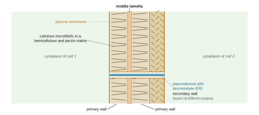
A growing plant cell is surrounded by a primary wall about 0.1 to 1 µm thick. Mechanically it behaves like a fiber-reinforced composite, comparable to reinforced concrete or a carbon-fiber panel. The reinforcing fibers are cellulose microfibrils; the matrix in which they are embedded is a hydrated gel of hemicelluloses and pectins, together with a small amount of structural protein. The proportions vary among species and tissues, but a typical dicot primary wall is roughly a quarter to a third cellulose, a quarter hemicellulose and a third pectin by dry mass, with most of its fresh mass being water.
Cellulose is an unbranched polymer of β-1,4-linked glucose. Because each glucose is rotated 180° relative to its neighbor, the chain is flat and ribbon-like, and parallel chains hydrogen-bond to one another to form crystalline microfibrils about 3 nm wide. Each microfibril is thought to contain about 18 to 24 chains. Cellulose is not made in the Golgi like other wall polysaccharides. It is synthesized at the plasma membrane by cellulose synthase complexes, rosette-shaped assemblies of CESA proteins visible in freeze-fracture electron micrographs. Each complex spins out several glucan chains at once, which crystallize immediately into a microfibril, and the complex moves through the membrane as the microfibril is laid down behind it. With a tensile strength along the fiber axis comparable to steel per unit cross-section, cellulose is the most abundant biopolymer on Earth.
Hemicelluloses are branched polysaccharides with a backbone of β-1,4-linked sugars that can hydrogen-bond to cellulose. In most dicots and non-grass monocots the main hemicellulose is xyloglucan, a glucan backbone decorated with xylose side chains; grasses use mainly glucuronoarabinoxylans and mixed-linkage glucans. The older textbook picture showed xyloglucan tethers coating microfibrils and linking neighboring fibers. Newer evidence, especially from solid-state nuclear magnetic resonance and enzyme studies, suggests that only a small fraction of xyloglucan is in tight contact with cellulose, at sites where microfibrils approach one another, so-called biomechanical hotspots. These junctions are thought to control how easily fibers can slide apart.
Pectins are a family of galacturonic acid-rich polysaccharides that form a hydrated gel. Homogalacturonan, a linear chain of galacturonic acid, is secreted in a methyl-esterified form and later de-esterified in the wall by pectin methylesterases; the freed carboxyl groups can then be cross-linked by calcium ions into an egg-box structure that stiffens the gel. Rhamnogalacturonan I carries arabinan and galactan side chains, and rhamnogalacturonan II is a complex molecule that is cross-linked into dimers by borate, which is why boron is an essential plant nutrient (lesson 6). The middle lamella, the layer that glues neighboring cells together, is especially rich in pectin, and fruit softening during ripening involves enzymes that break down pectin and loosen these junctions. The same chemistry explains why pectin sets jam.
Secondary walls and lignin
When a cell stops growing, it may deposit a secondary wall inside the primary wall. Secondary walls are thicker, often several micrometers, richer in cellulose and poorer in pectin, and they are laid down in layers in which microfibrils run at different angles, like the plies of plywood. In cells that provide support or conduct water, such as fibers, tracheids and vessel elements, the secondary wall is impregnated with lignin, a complex phenolic polymer that makes the wall hydrophobic, rigid and resistant to decay. Lignin is built from three main monolignols that give H (p-hydroxyphenyl), G (guaiacyl) and S (syringyl) units. Gymnosperm lignin is mostly G, while angiosperm wood contains both G and S units. Unusually for a biological polymer, lignin is assembled by radical coupling without a template, so its structure is irregular.
Lignin was one of the key innovations that let plants colonize land and grow tall. A water-conducting cell must withstand strong negative pressure (lesson 5) without collapsing inward, and lignified secondary walls provide that strength. Lignin also matters economically. It is the main obstacle in converting wood and straw into paper and into cellulosic biofuels, because it shields cellulose from enzymes, and plant breeders have produced crops with modified lignin, such as reduced-lignin alfalfa, to improve digestibility for livestock. Wood, cotton, linen and paper are all, at heart, products of the secondary wall.
| Polymer | Building blocks | Where it is made | Main role |
|---|---|---|---|
| Cellulose | β-1,4-glucose chains in crystalline microfibrils | Plasma membrane (CESA complexes) | Load-bearing fibers; resist tension |
| Xyloglucan | β-1,4-glucan backbone with xylose side chains | Golgi apparatus | Binds cellulose at junctions; controls fiber separation |
| Homogalacturonan | α-1,4-galacturonic acid | Golgi apparatus | Gel matrix; calcium cross-links stiffen the wall |
| Rhamnogalacturonan II | Highly branched pectic domain | Golgi apparatus | Borate cross-links; needed for normal growth |
| Lignin | Monolignols giving H, G and S units | Wall (radical polymerization) | Rigidity, waterproofing and defense in secondary walls |
| Extensins | Hydroxyproline-rich glycoproteins | Endoplasmic reticulum and Golgi | Cross-linked scaffolds, especially after wounding |
How walls grow: turgor, yielding and acid growth
A plant cell grows by taking up water into its vacuole while its wall yields and extends. Turgor pressure, typically 0.3 to 1 MPa or three to ten times atmospheric pressure, pushes outward on the wall, and the wall resists. Growth happens when the wall is loosened enough to creep irreversibly under that stress, and new material is added so the wall does not simply thin out. James Lockhart captured this in 1965 with a simple equation that is still the starting point for thinking about cell expansion. The relative growth rate equals a wall extensibility multiplied by the amount by which turgor exceeds a yield threshold:
Here \(P\) is turgor, \(Y\) is the yield threshold below which the wall does not extend, and \(\phi\) is the extensibility, which measures how readily the wall creeps once the threshold is passed. The equation shows that a cell can speed up its growth in two ways: by raising turgor, for example by accumulating solutes, or by loosening the wall, which raises \(\phi\) or lowers \(Y\). In practice, most fast changes in growth rate come from changes in the wall.
The best-understood mechanism of wall loosening is acid growth. In the 1970s, Robert Cleland, David Rayle and Achim Hager showed that auxin causes cells to pump protons into the wall, lowering the wall pH from about 6 to about 4.5 to 5, and that acidic buffers alone can make isolated walls extend. Daniel Cosgrove and colleagues identified the proteins responsible in the early 1990s and named them expansins. Expansins are not enzymes in the usual sense: they do not cut polysaccharides, but at acidic pH they let cellulose microfibrils slide relative to one another, probably by disrupting non-covalent bonds at the junctions between microfibrils and xyloglucan. Other proteins add to the effect. Xyloglucan endotransglucosylase/hydrolases (XTHs) cut and rejoin xyloglucan chains, and pectin-modifying enzymes alter the stiffness of the matrix. Lesson 7 returns to auxin and acid growth when it explains how a shoot bends toward light.
import numpy as np

# Lockhart model: relative growth rate r = phi * (P - Y) when P > Y, else 0
def rgr(P, Y, phi):
    return np.where(P > Y, phi * (P - Y), 0.0)

P = np.array([0.20, 0.30, 0.40, 0.50, 0.60, 0.70])   # turgor, MPa
Y = 0.30                                              # yield threshold, MPa
phi_neutral = 0.40      # extensibility at wall pH 6, per MPa per hour
phi_acid = 1.20         # extensibility after acidification (expansins active)

print(" P (MPa)  RGR pH 6 (%/h)  RGR pH 4.5 (%/h)")
for p, a, b in zip(P, rgr(P, Y, phi_neutral), rgr(P, Y, phi_acid)):
    print(f"  {p:4.2f}       {100*a:5.1f}           {100*b:5.1f}")

# hours for a cell to double in volume at P = 0.6 MPa
for label_, phi in (("pH 6", phi_neutral), ("pH 4.5", phi_acid)):
    r = phi * (0.6 - Y)
print(f"doubling time at {label_}: {np.log(2)/r:.1f} h")P (MPa) RGR pH 6 (%/h) RGR pH 4.5 (%/h) 0.20 0.0 0.0 0.30 0.0 0.0 0.40 4.0 12.0 0.50 8.0 24.0 0.60 12.0 36.0 0.70 16.0 48.0 doubling time at pH 6: 5.8 h doubling time at pH 4.5: 1.9 h
The output shows the logic of the model. Below the yield threshold, no growth occurs however long we wait. Above it, growth rises linearly with turgor. Tripling the extensibility, which is roughly what acidification can do in a responsive stem segment, triples the growth rate at any turgor above the threshold and shortens the doubling time from almost six hours to under two. Real cells are more complicated, because as a cell takes up water its turgor falls unless solutes are imported, and wall loosening is regulated over time, but the Lockhart picture is a useful frame for interpreting experiments with hormones, light and water stress.
Microfibril orientation controls cell shape
A wall that is reinforced equally in all directions expands equally in all directions, giving a sphere. Most plant cells instead elongate, because their microfibrils are laid down mainly in one direction, perpendicular to the axis of growth, like hoops around a barrel. Turgor pushes equally in all directions, but the hoops resist widening, so the cell grows longer. The direction in which cellulose is laid down is guided by cortical microtubules lying just beneath the plasma membrane: CESA complexes travel along tracks defined by the microtubules. Drugs that depolymerize microtubules, such as oryzalin, make elongating cells swell into balloons, and mutations that disturb microtubule organization give twisted or stunted organs.
There is a mechanical subtlety here. In a cylindrical cell under pressure, the stress in the wall around the circumference, the hoop stress, is twice the stress along the length. The wall must therefore be much stronger circumferentially simply to keep its shape, and the hoop-like orientation of microfibrils provides exactly that. Microtubules themselves appear to align with the direction of maximum stress in the wall, a feedback that may help tissues coordinate their growth.
import math

# Wall stress in a cylindrical cell: thin-walled pressure vessel formulas
P = 0.6e6        # turgor, Pa (0.6 MPa)
r = 20e-6        # cell radius, m (20 um)
t = 0.2e-6       # wall thickness, m (0.2 um)
hoop = P * r / t
axial = P * r / (2 * t)
print(f"hoop stress  = {hoop/1e6:.0f} MPa")
print(f"axial stress = {axial/1e6:.0f} MPa")
print(f"ratio hoop/axial = {hoop/axial:.1f}")
# compare: an inflated car wheel at 0.22 MPa, radius 0.3 m, wall 0.01 m
print(f"car wheel hoop stress = {0.22e6*0.3/0.01/1e6:.1f} MPa")hoop stress = 60 MPa axial stress = 30 MPa ratio hoop/axial = 2.0 car wheel hoop stress = 6.6 MPa
Although turgor is only a few times atmospheric pressure, the wall is so thin relative to the cell radius that the stress within it reaches tens of megapascals, about nine times that in the wall of a car tire. This is why primary walls need crystalline cellulose and why loosening the wall even slightly has such a large effect on growth.
Plastids: one organelle, many forms
All plastids descend from a single endosymbiotic cyanobacterium, and all of them in a given plant contain the same small genome, typically about 120 to 130 genes in flowering plants, encoding parts of the photosynthetic apparatus, ribosomal proteins and RNAs. Most plastid proteins, more than two thousand of them, are encoded in the nucleus and imported. What differs among plastid types is which nuclear genes are expressed and therefore what the plastid becomes. Proplastids in meristem cells are small and undifferentiated. In the light, they develop into chloroplasts with thylakoid membranes; in seedlings grown in darkness they become etioplasts, with a lattice-like prolamellar body that converts into thylakoids within hours of illumination. Amyloplasts store starch in roots, tubers and seeds, and in root cap cells they act as statoliths that sense gravity (lesson 9). Chromoplasts accumulate carotenoids and give the color of tomatoes, peppers and many flowers. Elaioplasts store lipids, and leucoplasts are colorless plastids that synthesize monoterpenes and other compounds.
Plastids do far more than photosynthesize. They make all of the cell’s fatty acids, many amino acids including the aromatic amino acids, purines, the carotenoid and chlorophyll pigments and precursors of several hormones, including abscisic acid, gibberellins, strigolactones and jasmonates. They also reduce nitrite and sulfate. The herbicide glyphosate works by blocking a plastid enzyme, EPSP synthase, in the pathway that makes aromatic amino acids, which animals do not have. Plastids interconvert as tissues develop: a green tomato turns red as its chloroplasts become chromoplasts, and a potato tuber exposed to light turns green as amyloplasts become chloroplasts.
The vacuole
The central vacuole is bounded by its own membrane, the tonoplast, and it is far more than a water balloon. By filling most of the cell with an inexpensive watery solution, the vacuole lets a plant make large cells cheaply; a leaf mesophyll cell may be 90 percent vacuole with a thin layer of cytoplasm pressed against the wall. The tonoplast carries two kinds of proton pumps, a V-type ATPase and a pyrophosphatase, that acidify the vacuole and energize the uptake of ions, sugars, organic acids and secondary metabolites. Many cells keep separate vacuoles for different jobs: lytic vacuoles that resemble animal lysosomes and contain hydrolytic enzymes, and protein storage vacuoles in seeds that hold the reserves used during germination.
Vacuoles store pigments such as anthocyanins, which change color with vacuolar pH, toxic compounds such as alkaloids and cyanogenic glycosides that would harm the cell’s own cytoplasm, and crystals of calcium oxalate. In CAM plants, the vacuole stores malic acid overnight (lesson 4), and in guard cells rapid exchange of potassium and chloride between vacuole and cytoplasm drives stomatal opening and closing (lesson 5). Aquaporins in the tonoplast allow water to move quickly into and out of the vacuole, buffering the cytoplasm against changes in water status.
Plasmodesmata and the symplast
Plasmodesmata are channels about 30 to 60 nm in diameter that cross the cell wall. Each is lined by plasma membrane that is continuous between the two cells, and through its center runs a narrow tube of compressed endoplasmic reticulum called the desmotubule. Molecules move through the cytoplasmic sleeve between the plasma membrane and the desmotubule. Primary plasmodesmata form during cytokinesis, where strands of endoplasmic reticulum are trapped in the forming cell plate; secondary plasmodesmata can be added later through existing walls.
Plasmodesmata divide the plant into two compartments. The symplast is the continuous cytoplasm of all cells connected by plasmodesmata; the apoplast is everything outside the plasma membranes, the walls and the water-filled spaces between cells, plus the dead conducting cells of the xylem. The size exclusion limit of a plasmodesma, the largest molecule that can pass by diffusion, is typically around 1 kDa in mature tissues, enough for sugars, amino acids and hormones, but plasmodesmata can dilate to allow transcription factors, small RNAs and viral movement proteins to pass. Plants regulate plasmodesmata by depositing callose, a β-1,3-glucan, in the wall around the neck of the channel, which narrows it; callose deposition is rapid after wounding and during pathogen attack. Lessons 6 and 7 show how the symplast and apoplast form alternative routes for water, nutrients and sugar, and lesson 11 shows how viruses exploit plasmodesmata to spread.
Worked problem
Reading a plasmodesmata experiment
A researcher loads fluorescent dyes of different sizes into one leaf epidermal cell and watches whether they spread to neighbors. A 0.5 kDa dye spreads within a minute; a 10 kDa labeled dextran stays in the injected cell. In a second leaf infected with a virus, the 10 kDa dextran spreads several cells away. What do these results show?
Show solution
- The small dye moves cell to cell without crossing membranes, so it travels through plasmodesmata in the symplast.
- The 10 kDa dextran is larger than the size exclusion limit of the uninfected tissue, so it cannot pass.
- In infected tissue, a molecule well above the normal limit now moves, so the plasmodesmata must have dilated.
- Viral movement proteins are known to increase the size exclusion limit; the result suggests the virus is gating plasmodesmata to spread its genome.
Answer: The experiment measures the size exclusion limit (between 0.5 and 10 kDa in healthy tissue) and shows that viral infection raises it. This classic approach, first used with tobacco mosaic virus movement protein, revealed that plasmodesmata are regulated channels rather than fixed pores.
Walls, plants and people
Cell walls make up most of the biomass on land, and almost every way people use plants touches them. Timber and paper are secondary walls; cotton is nearly pure cellulose from seed hairs; flax and hemp fibers are long lignified cells. Dietary fiber is plant cell wall material that human enzymes cannot digest. Ruminants can digest cellulose only with the help of microbes in the rumen, and lignin limits how much they can extract. Cellulosic biofuels and bio-based materials depend on separating cellulose from lignin and hemicellulose efficiently, and that remains costly. Understanding the wall at the molecular level is therefore not only basic science but also a route to better forages, stronger fibers and cheaper renewable materials. The next lesson moves up from the cell to the tissues and organs that walls make possible.
Common mistakes
MistakeThinking of the cell wall as a rigid, dead box.
InsteadThe primary wall is a dynamic composite that is loosened, extended and remodeled continually while the cell grows.
MistakeAssuming turgor alone determines how fast a cell grows.
InsteadIn the Lockhart model growth depends on the turgor above a yield threshold and on wall extensibility; most fast changes come from wall loosening.
MistakeDescribing expansins as enzymes that cut cellulose.
InsteadExpansins break no covalent bonds; at acidic pH they let microfibrils slide by disrupting non-covalent junctions.
MistakeSaying that cellulose is made in the Golgi like other wall polysaccharides.
InsteadCellulose is spun out by CESA complexes in the plasma membrane; hemicelluloses and pectins are made in the Golgi.
MistakeUsing chloroplast and plastid as synonyms.
InsteadChloroplasts are one type of plastid; amyloplasts, chromoplasts, etioplasts and others share the same genome but different roles.
MistakeTreating plasmodesmata as open holes of fixed size.
InsteadTheir size exclusion limit is regulated, for example by callose deposition, and can be widened by viral movement proteins.
Summary and key terms
- Plant cells cannot move, so plant form arises from the orientation of cell division and the direction of cell expansion.
- The primary wall is a composite of crystalline cellulose microfibrils in a hydrated matrix of hemicelluloses and pectins; secondary walls add more cellulose and, in many cells, lignin.
- Cellulose is made at the plasma membrane by moving CESA complexes guided by cortical microtubules; matrix polysaccharides are made in the Golgi.
- In the Lockhart model, relative growth rate equals extensibility times the excess of turgor over a yield threshold; acid growth and expansins raise extensibility.
- Hoop-like microfibrils resist widening and make cylindrical cells elongate; hoop stress is twice the axial stress.
- Plastids share one small genome but differentiate into chloroplasts, amyloplasts, chromoplasts and other forms with many biosynthetic roles.
- The vacuole provides cheap cell volume, turgor, storage and pH control; plasmodesmata join cells into a symplast with a regulated size exclusion limit.
- primary wall
- The thin, extensible wall laid down while a plant cell is growing.
- secondary wall
- A thicker wall deposited inside the primary wall after growth stops, often lignified.
- cellulose microfibril
- A crystalline bundle of parallel β-1,4-glucan chains that reinforces the wall.
- cellulose synthase complex
- A rosette of CESA proteins in the plasma membrane that synthesizes cellulose microfibrils.
- pectin
- A family of galacturonic acid-rich polysaccharides that form the gel matrix of the wall and the middle lamella.
- lignin
- A phenolic polymer that makes secondary walls rigid, waterproof and resistant to decay.
- expansin
- A wall protein that allows microfibrils to slide at acidic pH, loosening the wall.
- Lockhart equation
- A model in which relative growth rate equals wall extensibility times turgor in excess of a yield threshold.
- tonoplast
- The membrane surrounding the plant vacuole.
- plasmodesma
- A membrane-lined channel through the cell wall that connects the cytoplasm of neighboring cells.
- symplast
- The continuous cytoplasm of cells connected by plasmodesmata.
- apoplast
- The space outside plasma membranes, including cell walls, intercellular spaces and xylem.
Practice problems
Work each problem on paper first, then reveal the answer to check.
Name the three main classes of polysaccharide in a dicot primary wall and the role of each.
Show answer
Cellulose (load-bearing microfibrils), hemicelluloses such as xyloglucan (bind cellulose at junctions) and pectins (hydrated gel matrix, cross-linked by calcium).
Where is cellulose synthesized, and what guides the direction of deposition?
Show answer
At the plasma membrane by CESA complexes, which move along tracks defined by cortical microtubules.
In the Lockhart model, turgor is 0.5 MPa, the yield threshold is 0.3 MPa and extensibility is 0.5 per MPa per hour. What is the relative growth rate?
Show answer
0.5 × (0.5 − 0.3) = 0.10 per hour, or 10 percent per hour.
If turgor falls below the yield threshold, what happens to growth in the Lockhart model?
Show answer
Growth stops; the wall does not extend irreversibly below the threshold.
What is acid growth, and which proteins mediate it?
Show answer
Auxin-induced proton pumping acidifies the wall and activates expansins, which let microfibrils slide so the wall extends.
Why does a cylindrical cell with hoop-like microfibrils elongate rather than swell?
Show answer
The microfibrils resist circumferential expansion, so turgor-driven expansion occurs mainly along the long axis.
A cell has radius 15 µm, wall thickness 0.15 µm and turgor 0.5 MPa. What is the hoop stress?
Show answer
σ = Pr/t = 0.5 × 15/0.15 = 50 MPa.
Give three plastid types and a function of each.
Show answer
Chloroplasts (photosynthesis), amyloplasts (starch storage and gravity sensing), chromoplasts (carotenoid pigments in fruits and flowers).
Why is boron an essential nutrient for plants?
Show answer
It cross-links rhamnogalacturonan II in the wall; without it walls are weak and growth is abnormal.
How do plants close plasmodesmata after wounding?
Show answer
By depositing callose around the neck of the channel, reducing the size exclusion limit.
Lesson quiz · 5 questions
Score 60% or more to complete this lesson.
Create a free account to save your progress, take quizzes and earn a certificate.