
BIOL-207 · Lesson 4 of 12 · 32 min
DNA replication II: eukaryotes, telomeres and fidelity
Eukaryotic replication follows the same basic logic as bacterial replication: helicase opens the duplex, primers start new strands, polymerases extend them 5′ to 3′, and the lagging strand is made in fragments that are joined. But eukaryotic cells face three extra problems. Their genomes are far larger and are copied far more slowly, so one origin per chromosome would never be enough. Their chromosomes are linear, which creates a problem at the ends. And their DNA is wrapped in nucleosomes that must be taken apart and rebuilt. This lesson examines how cells solve those problems, and then asks how any cell copies billions of base pairs with fewer than one error per billion.
Many origins, once per cycle
A human cell replicates about 6.2 billion base pairs during S phase, which typically lasts around eight hours. Eukaryotic forks are slower than bacterial ones, moving at roughly 1 to 3 kb per minute, about 20 to 50 nucleotides per second, partly because they must deal with chromatin. A single origin per chromosome could not possibly finish in time. Instead, each human cell uses roughly 30,000 to 50,000 origins, spaced every 50 to 300 kb or so, which fire at different times during S phase.
Worked problem
Why eukaryotes need many origins
Human chromosome 1 is about 248 Mb. If forks move at 50 nt/s, how long would replication take from a single central origin? How many evenly spaced origins are needed to finish in 8 hours?
Show solution
- Single origin: each fork copies 124 Mb; 1.24 × 10⁸ / 50 = 2.48 × 10⁶ s ≈ 690 hours, or about 29 days.
- In 8 hours (28,800 s), one fork copies 50 × 28,800 = 1.44 × 10⁶ bp.
- Each origin, with two forks, can handle about 2.88 Mb.
- Origins needed ≈ 248 / 2.88 ≈ 86.
Answer: About a month from one origin, but fewer than a hundred evenly spaced origins could finish in 8 hours. Real cells use far more, often more than 2,000 on chromosome 1, because origins fire at different times and forks stall at obstacles, so extra origins provide speed and backup.
# Replication time for a human genome with different numbers of evenly spaced origins
genome = 6.2e9          # diploid bp
fork_speed = 40         # nt per second (illustrative)
for origins in (100, 1_000, 10_000, 30_000, 50_000):
    segment = genome / origins          # bp served by each origin (two forks)
    t_hours = (segment / 2) / fork_speed / 3600
print(f"{origins:6,d} origins: segment {segment/1e3:9,.0f} kb -> {t_hours:8.1f} h")100 origins: segment 62,000 kb -> 215.3 h 1,000 origins: segment 6,200 kb -> 21.5 h 10,000 origins: segment 620 kb -> 2.2 h 30,000 origins: segment 207 kb -> 0.7 h 50,000 origins: segment 124 kb -> 0.4 h
Having many origins creates a new danger: if any origin fired twice in one cell cycle, part of the genome would be copied twice, producing extra copies that destabilize chromosomes. Cells prevent this with a two-step licensing system. In G1, when activity of cyclin-dependent kinases (CDKs) is low, the origin recognition complex (ORC), together with the proteins Cdc6 and Cdt1, loads inactive double rings of the MCM helicase onto origins. This is licensing. In S phase, rising CDK and DDK activity activates the loaded helicases, adding partner proteins (Cdc45 and GINS) to form the active CMG helicase, and at the same time prevents any new MCM loading. Because loading and activation can happen only in different phases, each origin can fire at most once per cycle.
| Step | Cell-cycle phase | Key proteins | Result |
|---|---|---|---|
| Origin recognition | Throughout, bound in G1 | ORC | Marks potential origins |
| Licensing | G1 (low CDK activity) | Cdc6, Cdt1, MCM2–7 | Inactive MCM double rings loaded |
| Activation (firing) | S phase (high CDK and DDK) | Cdc45, GINS, MCM: the CMG helicase | Two forks move outward |
| Block to relicensing | S, G2 and M | CDK phosphorylation; geminin inhibits Cdt1 | No new loading until next G1 |
Eukaryotic polymerases
Eukaryotes divide the work among several polymerases. DNA polymerase α, bound to primase, makes a short RNA primer followed by a short stretch of DNA. Because polymerase α has low processivity and no proofreading, it hands off quickly. DNA polymerase ε then synthesizes most of the leading strand, and DNA polymerase δ synthesizes most of the lagging strand, both held on DNA by the sliding clamp PCNA (proliferating cell nuclear antigen), the eukaryotic equivalent of the bacterial β clamp. On the lagging strand, primers are removed by the nuclease FEN1 and by RNase H, polymerase δ fills the gaps, and DNA ligase I seals the nicks. Eukaryotes also have many specialized polymerases for repair and for copying past damaged bases.
| Function | E. coli | Eukaryotes |
|---|---|---|
| Initiator | DnaA | ORC |
| Replicative helicase | DnaB (moves 5′ to 3′) | CMG, built around MCM2–7 (moves 3′ to 5′) |
| Primase | DnaG | Primase subunits bound to Pol α |
| Leading-strand polymerase | Pol III | Pol ε |
| Lagging-strand polymerase | Pol III | Pol δ |
| Sliding clamp | β clamp (dimer) | PCNA (trimer) |
| Primer removal | Pol I 5′ to 3′ exonuclease | RNase H and FEN1 |
| Okazaki fragment size | 1,000–2,000 nt | 100–200 nt (about one nucleosome) |
| Origins | One (oriC) | Tens of thousands |
Chromatin and replication
Nucleosomes ahead of the fork must be disassembled to let the replisome pass, and both daughter duplexes must be repackaged behind it. Histone chaperones, such as FACT ahead of the fork and CAF-1 behind it, carry out this work. Old histones, carrying their chemical modifications, are distributed roughly equally to the two daughters, and newly made histones fill the gaps. Cells make a burst of new histones during S phase for exactly this reason. Because old histones carry marks that indicate whether a region was active or silent, this recycling helps daughter cells inherit patterns of gene expression, part of the epigenetic memory discussed in BIOL-201 lesson 12 and lesson 10 of this course. The size of eukaryotic Okazaki fragments, about the length of DNA in one nucleosome, appears to reflect how lagging-strand synthesis stops when polymerase δ meets a newly deposited nucleosome.
The end-replication problem
Linear chromosomes have a problem at their ends. On the lagging strand, the last Okazaki fragment needs an RNA primer, and when that primer is removed there is no upstream 3′ end from which a polymerase can fill the gap. In addition, the ends are processed by nucleases to leave a single-stranded 3′ overhang. As a result, each round of replication would shorten the chromosome by about 50 to 200 base pairs. Over many divisions, genes near the ends would be lost.
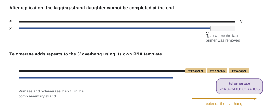
Cells solve this problem in two ways. First, chromosome ends are capped by telomeres, long arrays of a simple repeat that contain no genes, so losing some of the repeat does no immediate harm. In humans and other vertebrates, the repeat is TTAGGG, present in tandem copies stretching 5 to 15 kb at birth. Second, the enzyme telomerase can extend the 3′ end. Telomerase is a reverse transcriptase that carries its own RNA template (in humans, TERC) and a protein catalytic subunit (TERT). It binds the overhang, adds a TTAGGG repeat using its RNA as a template, moves along and adds another. The complementary strand is then filled in by primase and polymerase α. Elizabeth Blackburn, Carol Greider and Jack Szostak shared the 2009 Nobel Prize for discovering telomerase and how telomeres protect chromosomes.
The single-stranded overhang does not dangle free. A protein complex called shelterin binds telomeric DNA and folds the end back into a loop, the t-loop, in which the overhang invades the duplex region. This hides the end from the cell’s DNA damage machinery, which would otherwise treat it as a broken chromosome and try to join it to other ends.
Telomeres, aging and cancer
Most human somatic cells express little or no telomerase. Their telomeres shorten with each division. In the 1960s Leonard Hayflick showed that normal human fibroblasts in culture divide only a limited number of times, roughly 40 to 60, before entering a permanent non-dividing state called replicative senescence. Shortened telomeres are a major trigger of that limit. Germ cells and stem cells express telomerase and maintain their telomeres. Most cancer cells, by contrast, reactivate telomerase, about 85 to 90 percent of human tumors, and the rest use a recombination-based alternative, which is why telomerase is studied as a target for cancer therapy (BIOL-450).
Worked problem
How many divisions until senescence?
A cell line starts with telomeres averaging 10 kb and senesces when they average 5 kb. If each division shortens telomeres by 100 bp on average, how many divisions can the cells make? How would a loss of 70 bp per division change the answer?
Show solution
- Loss allowed = 10,000 − 5,000 = 5,000 bp.
- At 100 bp per division: 5,000 / 100 = 50 divisions.
- At 70 bp per division: 5,000 / 70 ≈ 71 divisions.
Answer: About 50 divisions at 100 bp per division, close to the Hayflick limit, and about 71 at 70 bp per division. The numbers are illustrative; real loss rates vary, and senescence is triggered by the shortest telomeres, not the average.
import numpy as np
rng = np.random.default_rng(207)

# 46 chromosomes x 2 ends = 92 telomeres; senescence when the SHORTEST falls below 3 kb.
start = rng.normal(9000, 1500, 92)      # bp, illustrative spread at the start
loss_mean, loss_sd = 100, 30            # bp lost per division per telomere
tel = start.copy()
div = 0
while tel.min() > 3000:
    tel -= np.clip(rng.normal(loss_mean, loss_sd, tel.size), 0, None)
    div += 1
print("starting mean telomere length:", round(start.mean()), "bp")
print("starting shortest telomere:   ", round(start.min()), "bp")
print("divisions until shortest < 3 kb:", div)
print("mean telomere length at senescence:", round(tel.mean()), "bp")starting mean telomere length: 9164 bp starting shortest telomere: 5418 bp divisions until shortest < 3 kb: 25 mean telomere length at senescence: 6656 bp
The simulation makes a subtle point. Senescence comes when the shortest telomere becomes critically short, long before the average reaches that length. This is why measurements of average telomere length in blood cells, sometimes marketed as tests of “biological age”, are only loosely related to an individual’s health. Telomere length varies widely between people of the same age and is influenced by genetics, and claims about lengthening telomeres with supplements are not supported by good evidence.
Fidelity: how accurate is replication?
Replication is astonishingly accurate. In E. coli and in human cells, the final error rate is roughly one mistake per billion to ten billion nucleotides copied (10⁻⁹ to 10⁻¹⁰). This accuracy comes from three layers of quality control, each multiplying the effect of the last.
The first layer is base selection. The polymerase active site fits a correctly paired Watson–Crick base pair snugly, and an incorrect pair has the wrong shape. The polymerase closes around a correct incoming dNTP but closes poorly around a wrong one, so incorporation is much slower. Base selection alone gives an error rate of about 1 in 10⁴ to 10⁵.
The second layer is proofreading. Replicative polymerases (Pol III in E. coli, Pol δ and ε in eukaryotes) have a 3′ to 5′ exonuclease activity. When a wrong nucleotide is added, the mismatched end pairs poorly, slips into the exonuclease site and is removed, and the polymerase tries again. Proofreading improves fidelity about 100-fold.
The third layer acts after the fork has passed. The mismatch repair system scans newly made DNA for mispaired bases, identifies which strand is new, removes a stretch of the new strand containing the error and resynthesizes it. In E. coli, the new strand is recognized because it is not yet methylated on adenines in GATC sequences; in eukaryotes, nicks in the new strand and the orientation of PCNA appear to mark it. Mismatch repair improves fidelity a further 100- to 1,000-fold. BIOL-201 lesson 10 described the mismatch repair proteins and the cancer syndrome (Lynch syndrome) caused by their loss.
Worked problem
How many errors per human cell division?
A human cell copies 6.2 × 10⁹ bp per division. Estimate the number of new errors per division for final error rates of 10⁻⁹ and 10⁻¹⁰. What would happen without proofreading and mismatch repair (error rate 10⁻⁵)?
Show solution
- At 10⁻⁹: 6.2 × 10⁹ × 10⁻⁹ ≈ 6 errors per division.
- At 10⁻¹⁰: 6.2 × 10⁹ × 10⁻¹⁰ ≈ 0.6 errors per division.
- At 10⁻⁵: 6.2 × 10⁹ × 10⁻⁵ = 62,000 errors per division.
Answer: Roughly one new error per cell division with all three layers working, but tens of thousands without proofreading and mismatch repair. That difference explains why mutations in polymerase proofreading domains or in mismatch repair genes strongly predispose to cancer.
genome = 6.2e9
layers = [("base selection only", 1e-5),
          ("+ proofreading (x100)", 1e-7),
          ("+ mismatch repair (x300)", 1e-7 / 300)]
for name, rate in layers:
print(f"{name:26s} error rate {rate:.1e} -> {genome * rate:12,.1f} errors per division")base selection only error rate 1.0e-05 -> 62,000.0 errors per division + proofreading (x100) error rate 1.0e-07 -> 620.0 errors per division + mismatch repair (x300) error rate 3.3e-10 -> 2.1 errors per division
These estimates connect directly to evolution and medicine. Errors that escape all three layers become mutations, the raw material of evolution (BIOL-108). In the human germ line, the per-generation mutation rate is about 1.2 × 10⁻⁸ per base pair, giving each child roughly 60 to 70 new mutations, most of them from the father because sperm precursors divide many more times than egg precursors (BIOL-201 lesson 10).
Replication stress
Forks frequently encounter obstacles: damaged bases, tightly bound proteins, transcribing RNA polymerases, unusual structures such as G-quadruplexes, and regions where both nucleotides of a pair are scarce. When a fork stalls, the cell activates checkpoint kinases (ATR and CHK1) that slow the cell cycle, suppress the firing of distant origins and stabilize the stalled fork until it can restart or be rescued by a fork coming from a adjacent origin. Some regions, called common fragile sites, replicate late and are prone to breakage under stress. Many cancer cells experience chronic replication stress because oncogenes drive them to replicate before they are ready, and drugs that inhibit ATR are being tested in clinical trials as a way to exploit this weakness.
Common mistakes
MistakeEukaryotic chromosomes each have a single origin, like bacteria.
InsteadEukaryotic chromosomes have many origins, tens of thousands per human genome.
MistakeAn origin can fire as often as needed during S phase.
InsteadLicensing in G1 and its block in S phase ensure each origin fires at most once per cycle.
MistakeTelomerase uses a DNA template.
InsteadTelomerase is a reverse transcriptase with its own RNA template.
MistakeProofreading is carried out by a 5′ to 3′ exonuclease.
InsteadProofreading uses the 3′ to 5′ exonuclease of replicative polymerases to remove the most recently added nucleotide.
MistakeAverage telomere length directly measures how long a person will live.
InsteadTelomere length varies widely and senescence depends on the shortest telomeres; it is a weak predictor for individuals.
Summary and key terms
- Eukaryotic forks are slow (tens of nucleotides per second), so tens of thousands of origins fire during S phase.
- Licensing (MCM loading by ORC, Cdc6 and Cdt1 in G1) and activation (CMG formation in S) ensure each origin fires once per cycle.
- Pol α–primase starts strands, Pol ε copies the leading strand and Pol δ the lagging strand, held by the PCNA clamp.
- Histone chaperones disassemble and reassemble nucleosomes, distributing old histones and their marks to both daughters.
- Linear chromosomes lose DNA at their ends each round; telomeres (TTAGGG repeats) buffer this loss and telomerase extends them.
- Most somatic cells lack telomerase and reach senescence; most cancers reactivate it.
- Base selection, proofreading and mismatch repair together give an error rate of about 10⁻⁹ to 10⁻¹⁰ per base.
- licensing
- Loading of inactive MCM helicases onto origins in G1, a prerequisite for firing in S phase.
- origin recognition complex (ORC)
- The protein complex that marks eukaryotic origins of replication.
- PCNA
- The eukaryotic sliding clamp that holds replicative polymerases on DNA.
- end-replication problem
- The inability to completely copy the 3′ ends of linear chromosomes.
- telomere
- A protective array of short repeats at the end of a linear chromosome.
- telomerase
- A reverse transcriptase that extends telomeres using its internal RNA template.
- shelterin
- The protein complex that binds and protects telomeres.
- replicative senescence
- Permanent arrest of cell division triggered by critically short telomeres.
- proofreading
- Removal of a misincorporated nucleotide by a polymerase’s 3′ to 5′ exonuclease.
- mismatch repair
- Post-replication correction of mispaired bases on the newly synthesized strand.
Practice problems
Work each problem on paper first, then reveal the answer to check.
A 100 Mb chromosome is copied with forks moving at 40 nt/s. How long would a single central origin take?
Show answer
Each fork copies 50 Mb: 5 × 10⁷ / 40 = 1.25 × 10⁶ s ≈ 347 hours.
Why can MCM helicases not be loaded during S phase?
Show answer
High CDK activity and geminin block Cdc6 and Cdt1, preventing licensing.
What would happen if an origin fired twice in one cycle?
Show answer
Part of the genome would be re-replicated, producing extra copies and genome instability.
Which eukaryotic polymerase synthesizes most of the lagging strand?
Show answer
DNA polymerase δ.
Why does the end-replication problem occur only on linear chromosomes?
Show answer
Circular chromosomes have no ends; there is always an upstream 3′ end to fill the last gap.
Write the human telomeric repeat on the G-rich strand.
Show answer
5′-TTAGGG-3′, repeated many times.
A cell line loses 80 bp per division from telomeres of 8 kb and senesces at 4 kb. How many divisions remain?
Show answer
4,000 / 80 = 50 divisions.
Why do most cancer cells reactivate telomerase?
Show answer
Unlimited division requires maintaining telomeres; otherwise cells would senesce or suffer chromosome fusions.
If base selection gives 10⁻⁵ errors and proofreading improves this 100-fold, what is the error rate before mismatch repair?
Show answer
10⁻⁷ per nucleotide.
How does E. coli mismatch repair know which strand is new?
Show answer
The new strand is temporarily unmethylated at GATC adenines.
Lesson quiz · 5 questions
Score 60% or more to complete this lesson.
Create a free account to save your progress, take quizzes and earn a certificate.