
BIOL-207 · Lesson 2 of 12 · 30 min
DNA and RNA structure and chromatin
The double helix is one of the most recognizable shapes in science, but its importance lies in details: which atoms bond to which, why the two strands run in opposite directions, why some regions melt more easily than others, and how two meters of it fit into a nucleus. This lesson builds DNA and RNA from their parts, explains the forces that hold the helix together, introduces the alternative forms DNA and RNA can take, and then follows DNA into chromatin, the packaged form in which it actually exists in eukaryotic cells. BIOL-107 introduced nucleotides as monomers; here we use that knowledge to understand how structure makes function possible.
Nucleotides
A nucleotide has three parts: a five-carbon sugar, a nitrogen-containing base attached to carbon 1′ of the sugar, and one or more phosphate groups attached to carbon 5′. The prime marks distinguish sugar carbons from the atoms of the base. In DNA the sugar is 2′-deoxyribose, which lacks an oxygen at the 2′ carbon; in RNA it is ribose, with a hydroxyl (–OH) group at 2′. That single oxygen matters a great deal: the 2′-OH makes RNA more chemically reactive and less stable, because it can attack the adjacent phosphate and break the backbone, especially in alkaline conditions. DNA’s greater stability suits its role as a long-term archive.
The bases come in two families. The purines, adenine (A) and guanine (G), have two fused rings. The pyrimidines, cytosine (C), thymine (T) and uracil (U), have a single ring. DNA uses A, G, C and T; RNA uses A, G, C and U. Thymine is uracil with an added methyl group, and one reason DNA uses T is that cytosine can lose an amino group spontaneously and become uracil. Because U does not belong in DNA, repair enzymes can recognize and remove it (BIOL-201 lesson 10). If DNA normally contained U, such damage would be invisible.
| Feature | DNA | RNA |
|---|---|---|
| Sugar | 2′-deoxyribose (H at 2′) | Ribose (OH at 2′) |
| Bases | A, G, C, T | A, G, C, U |
| Usual form | Double-stranded helix | Single-stranded, folding on itself |
| Chemical stability | High | Lower; 2′-OH can cleave the backbone |
| Main roles | Long-term storage of genetic information | Messenger, structural, catalytic and regulatory roles |
| Typical lifetime in a cell | The life of the cell and beyond | Minutes to days for most mRNAs |
Nucleotides join through phosphodiester bonds. The phosphate attached to the 5′ carbon of one nucleotide bonds to the 3′-OH of the previous one, building a sugar–phosphate backbone with the bases sticking out to one side. Every strand therefore has direction: a free 5′ phosphate at one end and a free 3′-OH at the other. By convention, sequences are written from 5′ to 3′, left to right. The direction matters because every polymerase in this course adds nucleotides only to a 3′ end, so all nucleic acid synthesis runs 5′ to 3′.
The double helix
In the Watson–Crick double helix, two strands wind around a common axis in a right-handed spiral. The backbones are on the outside and the bases stack in the middle, paired across the helix: A with T by two hydrogen bonds, and G with C by three. Each pair consists of one purine and one pyrimidine, so every pair has the same width, about 1.1 nanometers between sugar attachments, which keeps the helix uniform regardless of sequence. The two strands are antiparallel: one runs 5′ to 3′ upward while its partner runs 5′ to 3′ downward. Complementarity means that the sequence of one strand determines the sequence of the other.
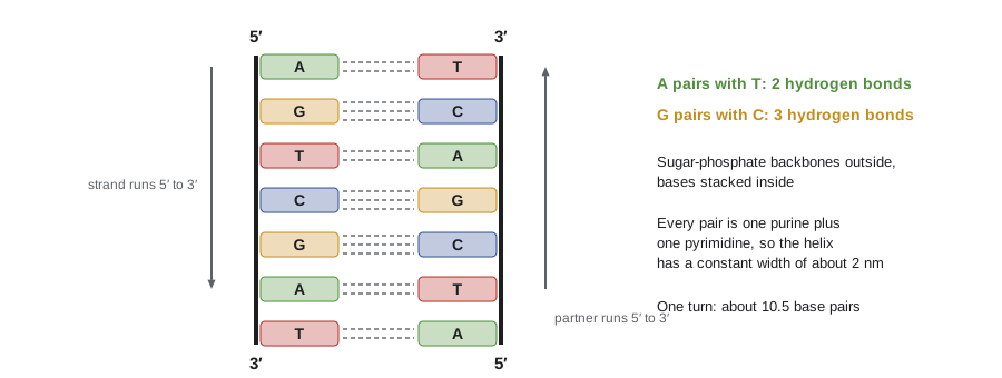
The common form of DNA in cells, called B-DNA, completes one turn about every 10.5 base pairs, and each base pair raises the helix by about 0.34 nm, so one turn is about 3.4 to 3.6 nm long. The helix diameter is about 2 nm. Because the sugars attach to the bases asymmetrically, the grooves between the backbones are of unequal size: a wide major groove and a narrow minor groove. Proteins that recognize specific DNA sequences, such as the transcription factors of lessons 9 and 10, usually read the edges of the base pairs exposed in the major groove without unwinding the helix.
Hydrogen bonds between bases are often described as what holds the helix together, but they are only part of the story. Base stacking, the attractive interactions between the flat faces of adjacent base pairs, contributes most of the stability. Water and positively charged ions also matter: the phosphate backbone carries a negative charge on every nucleotide, and without cations such as Na⁺ and Mg²⁺ to shield those charges, the two strands repel each other. This is why the stability of a duplex depends on salt concentration.
Writing and manipulating sequences
Because the strands are complementary and antiparallel, the partner of a strand written 5′-ATGCGT-3′ is 3′-TACGCA-5′, which, written in the conventional direction, is 5′-ACGCAT-3′. This operation, complementing and then reversing, is called taking the reverse complement, and it is one of the most common steps in sequence analysis. The code below defines it and checks one property: the reverse complement of the reverse complement is the original sequence.
COMP = str.maketrans("ACGTacgt", "TGCAtgca")

def revcomp(seq):
    """Reverse complement of a DNA sequence written 5' to 3'."""
    return seq.translate(COMP)[::-1]

def gc_content(seq):
    seq = seq.upper()
    return (seq.count("G") + seq.count("C")) / len(seq)

top = "ATGGCTAGCTTGACCGGAATTCTAA"
bottom = revcomp(top)
print("5'", top, "3'")
print("3'", bottom[::-1], "5'   (partner, aligned under the top strand)")
print("reverse complement, written 5'->3':", bottom)
print("round trip returns original:", revcomp(bottom) == top)
print(f"GC content: {gc_content(top):.1%}")5' ATGGCTAGCTTGACCGGAATTCTAA 3' 3' TACCGATCGAACTGGCCTTAAGATT 5' (partner, aligned under the top strand) reverse complement, written 5'->3': TTAGAATTCCGGTCAAGCTAGCCAT round trip returns original: True GC content: 44.0%
Melting and annealing
Heating DNA, or raising the pH, separates the strands, a process called denaturation or melting. When the solution cools slowly, complementary strands find each other and re-form the duplex, which is annealing or hybridization. The temperature at which half the DNA is single-stranded is the melting temperature, Tm. Because G–C pairs have three hydrogen bonds and stack more strongly, DNA with more G + C melts at a higher temperature. Melting can be followed by measuring absorbance of ultraviolet light at 260 nm, which rises by about 40 percent as the bases unstack, the hyperchromic effect.
Hybridization underlies most of the techniques in lessons 11 and 12, from PCR primers to probes and sequencing. For short synthetic DNA pieces called oligonucleotides, two simple estimates of Tm are widely used. The Wallace rule, suitable for oligos of about 14 to 20 nucleotides in standard salt, assigns 2 °C for each A or T and 4 °C for each G or C. A second formula, often used for longer sequences, adds a salt term:
In the second formula, \([\mathrm{Na}^+]\) is the molar sodium concentration and \(N\) is the length in nucleotides. Both are approximations; laboratories use more accurate nearest-neighbor models that account for which bases sit next to each other. They are good enough to compare candidate sequences and to see the effects of length, GC content and salt.
Worked problem
Estimating the melting temperature of a primer
A 20-nucleotide primer has the sequence 5′-AGCTTGACCGGAATTCAGGT-3′. Estimate its Tm by the Wallace rule and by the salt-adjusted formula at 50 mM Na⁺.
Show solution
- Count bases: A = 5, T = 5, G = 6, C = 4, so A + T = 10 and G + C = 10.
- Wallace rule: 2 × 10 + 4 × 10 = 60 °C.
- GC content = 10 / 20 = 50 percent; log₁₀(0.05) ≈ −1.30.
- Salt-adjusted: 81.5 + 16.6 × (−1.30) + 0.41 × 50 − 600/20 = 81.5 − 21.6 + 20.5 − 30.0 ≈ 50.4 °C.
Answer: About 60 °C by the Wallace rule and about 50 °C by the salt-adjusted formula. The two estimates differ by about 10 °C, a reminder that simple rules give rough values; primer design software uses nearest-neighbor thermodynamics.
import math

def tm_wallace(seq):
    s = seq.upper()
    return 2 * (s.count("A") + s.count("T")) + 4 * (s.count("G") + s.count("C"))

def tm_salt(seq, na=0.05):
    s = seq.upper()
    gc = 100 * (s.count("G") + s.count("C")) / len(s)
    return 81.5 + 16.6 * math.log10(na) + 0.41 * gc - 600 / len(s)

for primer in ["AGCTTGACCGGAATTCAGGT", "ATATTAAATTATATAATTTA", "GCGGCCGCGGCCGCGGCCGC"]:
    print(f"{primer}  Wallace {tm_wallace(primer):3d} C   salt-adjusted {tm_salt(primer):5.1f} C")
print()
for na in (0.01, 0.05, 0.2, 1.0):
print(f"[Na+] = {na:4.2f} M: Tm = {tm_salt('AGCTTGACCGGAATTCAGGT', na):5.1f} C")AGCTTGACCGGAATTCAGGT Wallace 60 C salt-adjusted 50.4 C ATATTAAATTATATAATTTA Wallace 40 C salt-adjusted 29.9 C GCGGCCGCGGCCGCGGCCGC Wallace 80 C salt-adjusted 70.9 C [Na+] = 0.01 M: Tm = 38.8 C [Na+] = 0.05 M: Tm = 50.4 C [Na+] = 0.20 M: Tm = 60.4 C [Na+] = 1.00 M: Tm = 72.0 C
The output shows the expected trends. An AT-rich oligo melts far below a GC-rich one of the same length, and raising the salt concentration raises Tm because cations shield the repulsion between the backbones.
Other DNA forms
B-DNA is not the only possibility. Under dehydrating conditions DNA adopts the A form, a wider, more compact right-handed helix; double-stranded RNA and DNA–RNA hybrids naturally take an A-like form because the 2′-OH prevents the sugar from adopting the B conformation. Z-DNA is a left-handed helix with a zigzag backbone that forms in some GC-rich sequences under strain; proteins that bind Z-DNA exist, and it may form transiently behind moving RNA polymerases. DNA can also form four-stranded G-quadruplexes in guanine-rich regions such as telomeres (lesson 4) and some promoters.
In cells, DNA is usually underwound, or negatively supercoiled. Picture a rubber band twisted until it coils on itself. Negative supercoiling stores energy that makes it easier to separate the strands for replication and transcription. Enzymes called topoisomerases add or remove supercoils by cutting and rejoining DNA. Type I topoisomerases cut one strand; type II cut both strands and pass another segment of the duplex through the break. Bacterial DNA gyrase, a type II enzyme that introduces negative supercoils, is the target of the fluoroquinolone antibiotics such as ciprofloxacin, and human topoisomerases are targets of several cancer drugs. Lesson 3 shows why replication needs topoisomerases.
RNA structure
Most RNA is single-stranded, but it folds back on itself wherever complementary stretches can pair, forming hairpins (stem-loops), bulges, internal loops and complex three-dimensional shapes. RNA also allows G–U pairs, called wobble pairs, which are important in transfer RNA (lesson 7). Folded RNAs can bind small molecules and even catalyze reactions. These catalytic RNAs, called ribozymes, include the peptidyl transferase center of the ribosome, the part that actually joins amino acids. The discovery of ribozymes by Thomas Cech and Sidney Altman, recognized with the 1989 Nobel Prize in Chemistry (Altman was born in Montréal and studied at McGill), supports the RNA world hypothesis: the idea that early life relied on RNA for both information storage and catalysis before DNA and proteins took over those roles.
| RNA class | Typical size | Role | Lesson |
|---|---|---|---|
| Messenger RNA (mRNA) | Hundreds to many thousands of nt | Carries coding information to ribosomes | 5–7 |
| Ribosomal RNA (rRNA) | 120 to about 5,000 nt | Structural and catalytic core of the ribosome | 7 |
| Transfer RNA (tRNA) | About 76 nt | Adapter that matches codons to amino acids | 7 |
| Small nuclear RNA (snRNA) | 100 to 200 nt | Part of the spliceosome | 6 |
| MicroRNA (miRNA) | About 22 nt | Represses target mRNAs | 10 |
| Long non-coding RNA (lncRNA) | More than 200 nt | Diverse regulatory roles, for example XIST | 10 |
Chromatin
In eukaryotes, DNA is wrapped around proteins to form chromatin. The basic unit is the nucleosome: about 147 base pairs of DNA wrapped 1.65 times around a core of eight histone proteins, two each of H2A, H2B, H3 and H4. Histones are rich in the positively charged amino acids lysine and arginine, which bind the negatively charged DNA backbone. Linker DNA, typically 20 to 60 bp, connects one nucleosome to the next, giving the “beads on a string” appearance seen in electron micrographs. A fifth histone, H1, binds the linker and helps fold the string into more compact fibers.
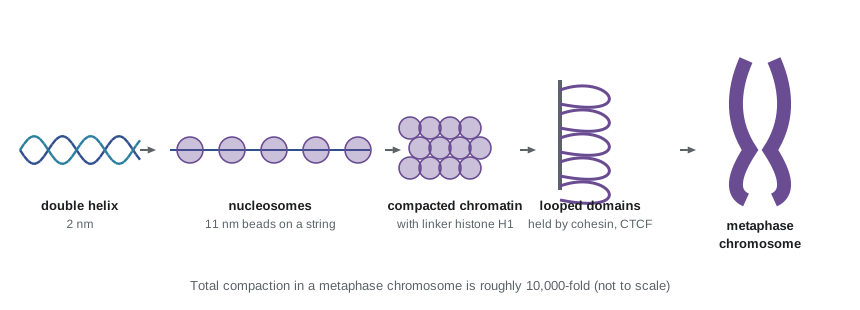
Beyond the nucleosome, chromatin folds into loops and domains. Modern methods that capture which parts of the genome touch each other, such as Hi-C, show that chromosomes are organized into topologically associating domains, regions of a few hundred kilobases that interact mostly within themselves, bounded by sites bound by the protein CTCF and held by cohesin rings. Lesson 10 explains how this folding brings enhancers close to the promoters they control. During mitosis, condensin complexes compact chromosomes further into the familiar metaphase shapes.
Chromatin is not uniform. Euchromatin is relatively open and contains most active genes. Heterochromatin is condensed and mostly silent; constitutive heterochromatin, such as the repetitive DNA around centromeres, is always condensed, while facultative heterochromatin, such as the inactive X chromosome in female mammals (BIOL-201 lesson 7), is condensed in some cells or at some times. The tails of histones stick out of the nucleosome and can be chemically modified by acetylation, methylation, phosphorylation and other marks. These modifications, together with DNA methylation, help determine whether a region is open or closed, a topic for lesson 10.
Worked problem
How much does the nucleosome compact DNA?
A nucleosome core plus linker uses about 200 bp of DNA and forms a bead about 11 nm long along the fiber. How much shorter is the DNA than if it were stretched out? How many nucleosomes are needed for the haploid human genome?
Show solution
- Stretched length of 200 bp = 200 × 0.34 nm = 68 nm.
- Packing ratio for the beads-on-a-string fiber = 68 / 11 ≈ 6.
- Nucleosomes per haploid genome ≈ 3.1 × 10⁹ / 200 ≈ 1.6 × 10⁷.
Answer: The nucleosome shortens DNA about six-fold, and a haploid genome needs about 15 million nucleosomes (about 30 million in a diploid cell). Higher levels of folding bring the total compaction in a metaphase chromosome to roughly 10,000-fold.
Prokaryotic genomes
Bacteria have no nucleus and no nucleosomes in the eukaryotic sense, but their DNA is still organized. The chromosome, usually a single circle, occupies a region called the nucleoid and is compacted by supercoiling and by abundant nucleoid-associated proteins such as HU and H-NS. Many bacteria also carry plasmids, small circular DNA molecules that replicate independently and often carry genes for antibiotic resistance. Plasmids spread between bacteria and are the basis of the cloning vectors of lesson 11. Archaea, the third domain of life, often have histone-like proteins related to eukaryotic histones, one of several clues that eukaryotes descended from an archaeal ancestor (BIOL-108).
Common mistakes
MistakeSequences can be written in either direction without ambiguity.
InsteadBy convention sequences are written 5′ to 3′; reversing a sequence without complementing it gives a different molecule.
MistakeHydrogen bonds are the main force holding the double helix together.
InsteadBase stacking contributes most of the stability; hydrogen bonds supply specificity.
MistakeRNA contains thymine and deoxyribose.
InsteadRNA contains uracil and ribose, with a 2′-OH group.
MistakeThe Wallace rule gives an accurate Tm for any DNA.
InsteadIt is a rough estimate for short oligos in standard salt; real Tm depends on sequence context, salt and concentration.
MistakeHistones bind specific DNA sequences.
InsteadHistone binding is largely sequence-independent, through attraction between positive histones and the negative DNA backbone.
Summary and key terms
- A nucleotide is a sugar, a base and phosphate; DNA uses deoxyribose and T, RNA uses ribose and U.
- Phosphodiester bonds link the 3′-OH of one nucleotide to the 5′ phosphate of the next, giving strands direction.
- B-DNA is a right-handed antiparallel double helix with A–T and G–C pairs, about 10.5 bp per turn, with major and minor grooves.
- Base stacking and salt-dependent shielding of the backbone stabilize the duplex; GC-rich DNA melts at higher temperatures.
- Simple formulas estimate oligo Tm; the reverse complement gives the sequence of the partner strand.
- RNA folds into hairpins and complex shapes; some RNAs are catalytic ribozymes.
- Eukaryotic DNA is packed into nucleosomes and higher-order chromatin; euchromatin is open, heterochromatin condensed.
- nucleotide
- A monomer of nucleic acid: a sugar, a nitrogenous base and one or more phosphates.
- phosphodiester bond
- The linkage joining the 3′ carbon of one nucleotide to the 5′ carbon of the next through a phosphate.
- antiparallel
- Running in opposite directions, as the two strands of a double helix do.
- major groove
- The wider groove of the double helix, where proteins most often read the bases.
- melting temperature (Tm)
- The temperature at which half of a DNA duplex has separated into single strands.
- reverse complement
- The sequence of the partner strand, written 5′ to 3′.
- supercoiling
- Twisting of a DNA duplex on itself; negative supercoiling eases strand separation.
- topoisomerase
- An enzyme that changes DNA supercoiling by cutting and rejoining strands.
- nucleosome
- About 147 bp of DNA wrapped around a core of eight histone proteins.
- heterochromatin
- Condensed, mostly transcriptionally silent chromatin.
Practice problems
Work each problem on paper first, then reveal the answer to check.
Write the reverse complement of 5′-GAATTCGG-3′.
Show answer
5′-CCGAATTC-3′.
Why does every DNA or RNA strand have a 5′ and a 3′ end?
Show answer
Because each phosphodiester bond links the 3′ carbon of one sugar to the 5′ carbon of the next, so the ends differ.
Which base pair has three hydrogen bonds?
Show answer
G–C.
Estimate the Wallace Tm of 5′-ATGCATGCATGCATGC-3′.
Show answer
8 A/T and 8 G/C: 2 × 8 + 4 × 8 = 48 °C.
Why does raising salt concentration raise the melting temperature?
Show answer
Cations shield the negative phosphates, reducing repulsion between the strands.
Why is DNA a better long-term storage molecule than RNA?
Show answer
It lacks the 2′-OH that makes RNA prone to backbone cleavage, and it is double-stranded, allowing repair from the partner strand.
What is the function of DNA gyrase, and why is it a good antibiotic target?
Show answer
It introduces negative supercoils in bacteria; bacteria need it, and it differs from human enzymes.
How many base pairs are in one turn of B-DNA?
Show answer
About 10.5.
Describe a nucleosome.
Show answer
About 147 bp of DNA wrapped 1.65 times around an octamer of histones H2A, H2B, H3 and H4.
Give one example of facultative heterochromatin.
Show answer
The inactive X chromosome in female mammals.
Lesson quiz · 5 questions
Score 60% or more to complete this lesson.
Create a free account to save your progress, take quizzes and earn a certificate.