
BIOL-207 · Lesson 1 of 12 · 34 min
The molecular basis of heredity
Genetics, as BIOL-201 showed, can be done without knowing what genes are made of. Mendel worked with abstract “factors”, and the first genetic maps were drawn decades before anyone knew the chemistry of a chromosome. Molecular biology asks the next question: what is the physical substance of heredity, and how does it store, copy and use information? This course follows that question from the experiments that identified DNA as the genetic material, through the machinery that copies DNA and reads it into RNA and protein, to the regulation that decides which genes are used when, and finally to the techniques that let us cut, copy, read and edit DNA in the laboratory.
This first lesson sets the scene. It reviews the experiments that led from “something in the chromosome” to DNA, introduces the central dogma and its exceptions, and practices the quantitative habits that run through the course: thinking in base pairs, kilobases and nucleotides per second, and checking claims against numbers. BIOL-107 introduced nucleic acids as one of the four classes of biological macromolecules, and BIOL-201 lesson 1 told the history of genetics in outline. Here we look more closely at how the key experiments were designed and why their conclusions were convincing.
Protein or nucleic acid?
By the 1920s it was clear that genes sat on chromosomes, and chemical analysis showed that chromosomes contain both protein and DNA. Most biologists assumed proteins were the genetic material. Proteins are built from 20 different amino acids and come in an enormous variety of shapes, while DNA, with only four kinds of nucleotide, looked too simple. A popular idea, the tetranucleotide hypothesis of Phoebus Levene, held that DNA was a monotonous repeat of the four bases in equal amounts, a structural scaffold rather than an information store. That idea turned out to be wrong, but it shaped thinking for two decades.
Griffith and transformation
In 1928 Frederick Griffith, a British medical officer studying pneumonia, worked with two strains of Streptococcus pneumoniae. The S strain made a polysaccharide capsule, formed smooth colonies and killed mice. The R strain lacked the capsule, formed rough colonies and was harmless. Heat-killed S bacteria were also harmless. But when Griffith injected mice with a mixture of live R bacteria and heat-killed S bacteria, many mice died, and he recovered live S bacteria from their blood. Something from the dead S cells had permanently changed the R cells into the virulent form, and the change was inherited by their descendants. Griffith called this transformation, without identifying the transforming substance.
Avery, MacLeod and McCarty
Oswald Avery, Colin MacLeod and Maclyn McCarty, working at the Rockefeller Institute in New York, spent more than a decade purifying the transforming principle. MacLeod was Canadian, trained at McGill. In 1944 they reported that the active material behaved chemically like DNA: it had the elemental composition of DNA, it was unaffected by enzymes that destroy protein (proteases) or RNA (ribonuclease), and it was destroyed by an enzyme that degrades DNA (deoxyribonuclease). The logic was elimination: if destroying a component abolishes activity, that component is needed; if destroying it leaves activity intact, it is not.
| Treatment of the S-strain extract | Transformation of R cells? | Conclusion |
|---|---|---|
| None | Yes | The extract contains the transforming principle |
| Protease (destroys protein) | Yes | Protein is not required |
| Ribonuclease (destroys RNA) | Yes | RNA is not required |
| Deoxyribonuclease (destroys DNA) | No | DNA is required |
| Polysaccharide-degrading enzyme | Yes | The capsule itself is not the agent |
Many scientists were not fully persuaded. Critics argued that tiny amounts of contaminating protein might be responsible, and that bacterial transformation might be a special case. The evidence was strong, but the conclusion was revolutionary, and revolutions in science usually need more than one line of evidence.
Hershey and Chase
The second line came in 1952 from Alfred Hershey and Martha Chase at Cold Spring Harbor, using bacteriophage T2, a virus that infects Escherichia coli. A phage is little more than DNA packed inside a protein coat. When it infects a cell, new phages appear inside after about 20 minutes. Hershey and Chase asked which part of the phage enters the cell and directs the production of new phages. Their insight was to label the two components separately with radioactive isotopes: protein contains sulfur (in the amino acids cysteine and methionine) but almost no phosphorus, while DNA contains phosphorus in its backbone but no sulfur.
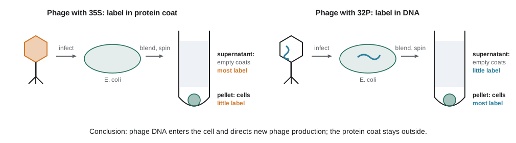
They grew one batch of phages with ³⁵S, labeling the protein coats, and another with ³²P, labeling the DNA. Each batch infected unlabeled bacteria. After a few minutes they agitated the mixtures in a kitchen blender to shear the phage coats off the bacterial surfaces, then centrifuged. The heavier bacterial cells formed a pellet; the lighter phage coats stayed in the liquid above (the supernatant). Most of the ³²P ended up in the pellet with the cells, and that label appeared in the next generation of phages. Most of the ³⁵S stayed in the supernatant with the empty coats.
Worked problem
Reading the Hershey–Chase numbers
In an experiment like Hershey and Chase’s, a researcher measures 1,000,000 counts per minute (cpm) of ³²P in the infected culture. After blending and centrifuging, 700,000 cpm are in the pellet and 300,000 cpm in the supernatant. For ³⁵S, 1,000,000 cpm total gives 200,000 in the pellet and 800,000 in the supernatant. What fraction of each label entered the cells, and what is the conclusion?
Show solution
- Fraction of ³²P (DNA) in the pellet: 700,000 / 1,000,000 = 0.70.
- Fraction of ³⁵S (protein) in the pellet: 200,000 / 1,000,000 = 0.20.
- The ³⁵S in the pellet comes mainly from coats still stuck to cells after incomplete blending, so it is a background level, not evidence that protein entered.
- Most DNA entered the cells; most protein did not.
Answer: About 70 percent of the DNA label entered the cells compared with about 20 percent of the protein label (largely incompletely removed coats), consistent with DNA being the material injected into the cell. The numbers are illustrative but close to the original pattern.
Because blending was imperfect, neither separation was clean, and Hershey and Chase themselves were cautious. But the experiment was simple, the result was easy to repeat, and coming after Avery’s work it settled the question for most biologists.
Chargaff’s rules and the double helix
Meanwhile Erwin Chargaff, at Columbia University, measured the base composition of DNA from many species. Two observations demolished the tetranucleotide hypothesis. First, base composition varies between species, which is what an information-carrying molecule should do. Second, within any double-stranded DNA the amount of adenine equals the amount of thymine, and the amount of guanine equals the amount of cytosine. These became known as Chargaff’s rules.
The structure that explained these rules was published in April 1953 by James Watson and Francis Crick at Cambridge. It drew critically on X-ray diffraction images made at King’s College London by Rosalind Franklin and her student Raymond Gosling, especially the image known as Photograph 51, and on measurements from Franklin’s unpublished report. Maurice Wilkins, also at King’s, shared the image with Watson without Franklin’s knowledge. The same issue of Nature carried papers by Wilkins and colleagues and by Franklin and Gosling supporting the model. Watson, Crick and Wilkins received the 1962 Nobel Prize; Franklin had died of ovarian cancer in 1958 at 37, and the prize is not awarded posthumously. Historians now agree that her data and analysis were essential to the discovery, and the story is often used to discuss credit and recognition in science.
Lesson 2 examines the structure in detail. The point for now is that the double helix immediately suggested how DNA could be copied: each strand carries the information to specify its partner. Watson and Crick ended their paper with a famously understated sentence noting that the specific pairing “immediately suggests a possible copying mechanism”. Lesson 3 shows how that mechanism was demonstrated by Meselson and Stahl in 1958.
Worked problem
Using Chargaff’s rules
A sample of double-stranded DNA contains 22 percent guanine. What are the percentages of the other three bases?
Show solution
- G = C, so cytosine is also 22 percent.
- G + C = 44 percent, so A + T = 100 − 44 = 56 percent.
- A = T, so each is 56 / 2 = 28 percent.
Answer: A = 28 percent, T = 28 percent, G = 22 percent, C = 22 percent. The GC content of this DNA is 44 percent.
The code below shows Chargaff’s rules emerging from base pairing. It generates a random single strand with a deliberately uneven composition, builds the complementary strand, and counts bases in the single strand and in the duplex. The single strand need not obey the rules; the double-stranded molecule always does.
import random
random.seed(207)

pair = {"A": "T", "T": "A", "G": "C", "C": "G"}
weights = {"A": 0.35, "C": 0.15, "G": 0.20, "T": 0.30}   # deliberately uneven
strand = "".join(random.choices(list(weights), weights=list(weights.values()), k=10_000))
partner = "".join(pair[b] for b in strand)

def pct(seq):
    return {b: round(100 * seq.count(b) / len(seq), 1) for b in "ACGT"}

print("one strand:      ", pct(strand))
print("double-stranded: ", pct(strand + partner))
duplex = strand + partner
print("A = T?", duplex.count("A") == duplex.count("T"), " G = C?", duplex.count("G") == duplex.count("C"))one strand: {'A': 35.3, 'C': 15.0, 'G': 19.8, 'T': 29.9}
double-stranded: {'A': 32.6, 'C': 17.4, 'G': 17.4, 'T': 32.6}
A = T? True G = C? TrueThe central dogma
In 1958 Francis Crick stated what he called the central dogma of molecular biology: information, once it has passed into protein, cannot get out again. The familiar summary is that DNA is copied into DNA (replication), DNA is transcribed into RNA (transcription) and RNA is translated into protein (translation). Crick’s original formulation was about which transfers of sequence information are possible, not about the importance of each molecule, and it allowed for transfers he thought might occur occasionally.
Several of the special transfers were later found. Retroviruses such as HIV carry RNA genomes and use the enzyme reverse transcriptase to copy RNA into DNA, discovered independently by Howard Temin and David Baltimore in 1970. Many RNA viruses, including influenza and the coronaviruses, copy RNA directly into RNA. Telomerase, which maintains the ends of our chromosomes (lesson 4), is a reverse transcriptase that carries its own RNA template. And a large fraction of the human genome consists of retrotransposons, sequences that spread by being transcribed and then reverse transcribed back into DNA (BIOL-201 lesson 10). What has never been found is a mechanism that copies the sequence of a protein back into nucleic acid. Prions, the infectious misfolded proteins behind diseases such as chronic wasting disease in deer and elk, transmit a shape, not a sequence, and so do not break the rule.
| Transfer | Enzyme | Where it happens |
|---|---|---|
| DNA to DNA (replication) | DNA polymerase | All cellular life; DNA viruses |
| DNA to RNA (transcription) | RNA polymerase | All cellular life |
| RNA to protein (translation) | Ribosome | All cellular life |
| RNA to DNA (reverse transcription) | Reverse transcriptase | Retroviruses, retrotransposons, telomerase |
| RNA to RNA (RNA replication) | RNA-dependent RNA polymerase | Many RNA viruses |
Genes, genomes and scale
Molecular biology is full of numbers, and getting a feel for them is part of understanding the field. DNA length is measured in base pairs (bp), kilobase pairs (kb, 1,000 bp) and megabase pairs (Mb, one million bp). The E. coli genome is about 4.6 Mb and encodes roughly 4,300 proteins; the human haploid genome is about 3.1 billion bp and contains roughly 20,000 protein-coding genes, which together occupy only about 1 to 2 percent of the sequence. One base pair adds about 0.34 nanometers of length to the double helix, so the DNA in a single human cell, stretched out, would be about two meters long, packed into a nucleus a few micrometers across. Lesson 2 shows how chromatin achieves that packing.
# Lengths of DNA molecules from base pair counts (0.34 nm per base pair)
genomes = {
    "phage lambda": 48_502,
    "E. coli": 4_641_652,
    "baker's yeast (haploid)": 12_100_000,
    "human (haploid)": 3_100_000_000,
}
NM_PER_BP = 0.34
for name, bp in genomes.items():
    length_m = bp * NM_PER_BP * 1e-9
    if length_m < 1e-3:
        print(f"{name:24s} {bp:>15,d} bp  ->  {length_m * 1e6:8.1f} micrometres")
    else:
        print(f"{name:24s} {bp:>15,d} bp  ->  {length_m * 1e3:8.1f} millimetres")
diploid_m = 2 * genomes["human (haploid)"] * NM_PER_BP * 1e-9
print(f"\nDNA in one diploid human cell: about {diploid_m:.1f} m")phage lambda 48,502 bp -> 16.5 micrometers E. coli 4,641,652 bp -> 1.6 millimeters baker's yeast (haploid) 12,100,000 bp -> 4.1 millimeters human (haploid) 3,100,000,000 bp -> 1054.0 millimeters DNA in one diploid human cell: about 2.1 m
Note the units in the output: the code converts to micrometers and millimeters because those are easier to picture than raw meters. A useful habit is to estimate before calculating. The human genome is about 3 × 10⁹ bp; multiplying by 3.4 × 10⁻¹⁰ m gives about 1 m per haploid set, so roughly 2 m per diploid cell, which matches the output.
Worked problem
How much DNA is there in your body?
An adult human body contains roughly 3 × 10¹³ cells (most of them red blood cells, which have no nucleus). Suppose about 1 × 10¹³ cells contain a full diploid genome of about 2 m of DNA. Estimate the total length of DNA, and compare it with the distance from Earth to the Sun (about 1.5 × 10¹¹ m).
Show solution
- Total length ≈ 1 × 10¹³ cells × 2 m per cell = 2 × 10¹³ m.
- Ratio to the Earth–Sun distance: 2 × 10¹³ / 1.5 × 10¹¹ ≈ 130.
Answer: About 2 × 10¹³ m, enough to stretch from Earth to the Sun and back more than 60 times. The cell counts are rough estimates, so the answer is an order of magnitude, which is all such a question needs.
Model organisms and tools
The founding experiments of molecular biology used bacteria and their viruses because they grow fast, in enormous numbers, on simple media. E. coli divides every 20 minutes or so under good conditions, so a single cell can become billions overnight. Phages made it possible to study gene structure at the level of individual nucleotides decades before DNA could be sequenced. Later, baker’s yeast (Saccharomyces cerevisiae) became the workhorse for eukaryotic molecular biology, joined by the fruit fly, the nematode Caenorhabditis elegans, the plant Arabidopsis thaliana, the zebrafish, the mouse and cultured human cells. Throughout this course, examples come from whichever organism revealed a mechanism most clearly, with the understanding that the core machinery of replication, transcription and translation is shared across life.
Canadian laboratories contributed to several stories in this course. Oswald Avery was himself born in Halifax, Nova Scotia, and MacLeod grew up in Prince Edward Island and Saskatchewan. Later, in the 1970s and 1980s, groups in Canada helped identify genes for cystic fibrosis and Duchenne muscular dystrophy (BIOL-201), and Canadian scientists contributed to the lipid nanoparticle technology used in mRNA vaccines, which lesson 12 discusses.
How to read this course
Each lesson moves from mechanism to quantity. You will meet the molecules involved, see how experiments revealed their roles, and then calculate: replication times, error rates, melting temperatures, binding fractions, amplification factors and sequencing coverage. Short Python programs, written with only the standard library or numpy, model these processes and handle DNA sequences as strings of letters. You do not need to be a programmer to follow them; each one is explained in the surrounding text, and the output is shown. If you want to experiment, change a number and predict the result before running the code again.
Common mistakes
MistakeGriffith showed that DNA is the genetic material.
InsteadGriffith discovered transformation; Avery, MacLeod and McCarty identified the transforming principle as DNA in 1944.
MistakeIn the Hershey–Chase experiment, ³²P labeled the phage proteins.
Instead³²P labels DNA (phosphate backbone); ³⁵S labels protein (sulfur in cysteine and methionine).
MistakeChargaff’s rules mean every DNA has 25 percent of each base.
InsteadThey mean A = T and G = C in double-stranded DNA; the proportion of G + C varies between species.
MistakeThe central dogma says information can never flow from RNA to DNA.
InsteadReverse transcription is allowed; the dogma forbids transfer of sequence information out of protein.
MistakeWatson and Crick determined the structure using their own X-ray images.
InsteadThey built a model using X-ray data from Rosalind Franklin and Raymond Gosling and from Maurice Wilkins, together with Chargaff’s rules.
Summary and key terms
- Griffith (1928) discovered transformation of harmless R pneumococci into virulent S pneumococci by heat-killed S cells.
- Avery, MacLeod and McCarty (1944) used enzymes to show that the transforming principle was DNA, not protein or RNA.
- Hershey and Chase (1952) showed that phage DNA (³²P), not protein (³⁵S), enters bacteria and directs new phage production.
- Chargaff’s rules (A = T, G = C) and variable base composition showed DNA could carry information and pointed to base pairing.
- The double helix (1953) was built on X-ray data from Franklin, Gosling and Wilkins; Franklin’s contribution was essential.
- The central dogma describes information flow from DNA to RNA to protein; reverse transcription and RNA replication are permitted exceptions.
- Genome sizes range from tens of kilobases in phages to billions of base pairs in humans; each base pair adds 0.34 nm of length.
- transformation
- The heritable change of a cell’s properties by uptake of DNA from its surroundings.
- transforming principle
- The substance in heat-killed S pneumococci that transformed R cells, identified as DNA.
- bacteriophage
- A virus that infects bacteria, often called a phage.
- radioactive labeling
- Marking a molecule with a radioactive isotope so it can be traced.
- Chargaff’s rules
- In double-stranded DNA, the amount of adenine equals thymine and guanine equals cytosine.
- central dogma
- Crick’s statement that sequence information flows from nucleic acid to protein but not back out of protein.
- reverse transcriptase
- An enzyme that copies RNA into DNA.
- base pair (bp)
- Two complementary nucleotides on opposite strands; also the unit of DNA length.
- genome
- The complete set of genetic material of an organism.
- model organism
- A species studied in depth because it is easy to grow and manipulate, with findings that apply more widely.
Practice problems
Work each problem on paper first, then reveal the answer to check.
Why did Griffith’s mixture of live R and heat-killed S bacteria kill mice when neither did alone?
Show answer
Material from the dead S cells transformed some R cells into live, capsule-forming, virulent S cells.
In Avery’s experiments, what result showed that RNA was not the transforming principle?
Show answer
Treating the extract with ribonuclease did not abolish transformation.
Why did Hershey and Chase use sulfur and phosphorus isotopes?
Show answer
Protein contains sulfur but little phosphorus; DNA contains phosphorus but no sulfur, so each label marks one component.
A double-stranded DNA is 31 percent adenine. Give the percentages of T, G and C.
Show answer
T = 31, G = 19, C = 19 percent.
A single-stranded viral DNA is 31 percent adenine and 18 percent thymine. Do Chargaff’s rules apply? Why?
Show answer
No; the rules depend on base pairing between two strands.
Name two information transfers that the central dogma allows but that are less common than replication, transcription and translation.
Show answer
Reverse transcription (RNA to DNA) and RNA replication (RNA to RNA).
Why are prions not an exception to the central dogma?
Show answer
They transmit a protein shape, not a nucleotide or amino acid sequence.
Estimate the length of the 4.6 Mb E. coli chromosome.
Show answer
4.6 × 10⁶ × 0.34 nm ≈ 1.6 mm.
Why was the tetranucleotide hypothesis important to overturn?
Show answer
It suggested DNA was too uniform to carry information, which led researchers to assume genes were proteins.
What made bacteria and phages good organisms for early molecular biology?
Show answer
They grow very quickly in huge numbers on simple media and have small genomes.
Lesson quiz · 5 questions
Score 60% or more to complete this lesson.
Create a free account to save your progress, take quizzes and earn a certificate.