
BIOL-207 · Lesson 10 of 12 · 31 min
Gene regulation in eukaryotes
Every cell in your body carries essentially the same genome, yet a neuron, a liver cell and a white blood cell look and behave completely differently. The difference lies in which genes each cell expresses and how strongly. Eukaryotic gene regulation is more elaborate than the bacterial switches of lesson 9, for several reasons: DNA is packaged in chromatin, which must be opened before genes can be read; regulatory sequences can lie tens or hundreds of kilobases from the genes they control; many transcription factors act in combination; and RNA processing, export, translation and decay each add further layers. This lesson works through those layers, from chromatin to microRNAs, with quantitative models of how regulators bind their targets.
Levels of control
Expression of a eukaryotic gene can be regulated at each step from DNA to active protein. Transcription remains the most important control point for most genes, because it is economical to avoid making RNA that will not be used. But the other levels allow faster or more localized responses.
| Level | Examples of mechanisms | Lesson |
|---|---|---|
| Chromatin | Histone modifications, nucleosome remodeling, DNA methylation | 10 (and BIOL-201 lesson 12) |
| Transcription initiation | Activators and repressors at enhancers; Mediator; general factors | 6, 10 |
| Elongation | Release from promoter-proximal pausing | 6 |
| RNA processing | Alternative splicing, alternative polyadenylation | 6 |
| Export and localization | Selective export; mRNAs targeted to parts of the cell | 6 |
| mRNA stability | AU-rich elements, microRNAs, poly(A) shortening | 10 |
| Translation | Control of initiation factors; RNA-binding proteins; microRNAs | 7, 10 |
| Protein activity and stability | Phosphorylation, ubiquitination, cleavage | 8 |
Chromatin as a barrier and a signal
Nucleosomes (lesson 2) cover most of the genome, and DNA wrapped around a histone octamer is largely inaccessible to transcription factors and RNA polymerase. Active promoters and enhancers are typically nucleosome-depleted regions where the DNA is open. Several kinds of enzyme control this openness. ATP-dependent chromatin remodelers, such as the SWI/SNF complex, slide nucleosomes along DNA, eject them or exchange histone variants. Histone-modifying enzymes add or remove chemical marks on the histone tails. And DNA methyltransferases add methyl groups to cytosines.
Histone acetylation, added by histone acetyltransferases (HATs) and removed by histone deacetylases (HDACs), neutralizes the positive charge of lysines, loosening the contact between histones and DNA and creating binding sites for proteins with bromodomains that promote transcription. Acetylation is therefore generally associated with active chromatin. Histone methylation is more nuanced: its effect depends on which residue is methylated and how many methyl groups are added. Trimethylation of lysine 4 on histone H3 (written H3K4me3) marks active promoters; H3K27me3, added by Polycomb complexes, marks repressed developmental genes; H3K9me3 marks constitutive heterochromatin and is bound by heterochromatin protein 1 (HP1).
| Mark | Usual location | Associated state |
|---|---|---|
| H3K27ac (acetylation) | Active enhancers and promoters | Active |
| H3K4me3 | Promoters of active genes | Active |
| H3K4me1 | Enhancers (active or poised) | Primed |
| H3K36me3 | Bodies of transcribed genes | Transcribed |
| H3K27me3 | Developmental genes kept off by Polycomb | Repressed, reversible |
| H3K9me3 | Repeats, centromeres, some genes | Heterochromatin, stably silent |
| DNA methylation at CpG | Repeats; silenced promoters; gene bodies | Promoter methylation silences |
In mammals, DNA methylation occurs mostly at cytosines followed by guanine, the CpG dinucleotide. About 70 to 80 percent of CpGs in the human genome are methylated, but CpG islands, regions of about 1 kb with a high density of CpGs, are found at the promoters of most genes and are usually unmethylated when the gene is active. Methylation of a CpG island promoter typically silences the gene, by blocking factor binding and recruiting proteins that bind methylated DNA and attract HDACs. Methylation patterns are copied during replication by DNMT1, which recognizes hemimethylated DNA. BIOL-201 lesson 12 discussed the role of methylation in genomic imprinting and X inactivation; cancer cells frequently show abnormal methylation, with tumor suppressor genes silenced by promoter methylation.
import random
random.seed(207)

def cpg_stats(seq):
    """Observed/expected CpG ratio, a standard test for CpG islands."""
    c, g, cg = seq.count("C"), seq.count("G"), seq.count("CG")
    gc = (c + g) / len(seq)
    expected = c * g / len(seq)
    return gc, cg / expected if expected else 0

# Bulk genome: CpG depleted (methylated C mutates to T over evolution)
import re
raw = "".join(random.choices("ACGT", weights=[0.30, 0.20, 0.20, 0.30], k=4000))
bulk = re.sub("CG", lambda m: "TG" if random.random() < 0.8 else "CG", raw)   # lose most CpGs
# A CpG island: GC-rich and not depleted
island = "".join(random.choices("ACGT", weights=[0.17, 0.33, 0.33, 0.17], k=1000))
for name, s in (("bulk DNA", bulk), ("CpG island", island)):
    gc, oe = cpg_stats(s)
    print(f"{name:11s} GC = {gc:.0%}  CpG observed/expected = {oe:.2f}")
print("Typical CpG island criteria: GC > 50% and observed/expected > 0.6 over 200+ bp")bulk DNA GC = 37% CpG observed/expected = 0.28 CpG island GC = 67% CpG observed/expected = 1.09 Typical CpG island criteria: GC > 50% and observed/expected > 0.6 over 200+ bp
The bulk of the human genome is depleted of CpG, at about a quarter of the frequency expected from its base composition. The reason is mutation: methylated cytosine can spontaneously lose an amino group and become thymine, a change that is harder to repair than conversion of ordinary cytosine to uracil. Over evolutionary time, methylated CpGs have been steadily converted to TpG. CpG islands escape this loss because they are kept unmethylated in the germ line.
Transcription factors
Transcription factors are proteins that bind specific DNA sequences and influence transcription. Humans have about 1,600. Most have a modular structure: a DNA-binding domain, which recognizes a short sequence of about 6 to 12 bp, and one or more activation or repression domains, which interact with co-activators, co-repressors, Mediator and chromatin modifiers. Classic experiments in yeast showed that the DNA-binding domain of one factor can be fused to the activation domain of another to make a working hybrid, demonstrating that the two functions are separable.
| DNA-binding domain family | Structural feature | Example |
|---|---|---|
| Homeodomain | Helix-turn-helix of 60 amino acids | HOX proteins that pattern the body axis |
| Zinc finger | Small folds stabilized by zinc ions; often many in a row | CTCF; nuclear hormone receptors |
| Basic leucine zipper (bZIP) | Basic DNA-binding region and a coiled-coil dimerization zipper | AP-1 (Fos and Jun) |
| Basic helix-loop-helix (bHLH) | Basic region plus two helices for dimerization | MyoD, which drives muscle differentiation |
Binding: a quantitative view
Whether a factor occupies its site depends on its concentration and its affinity, measured by the dissociation constant Kd, the concentration at which half the sites are occupied. The fraction of sites bound, θ, follows the same equation as ligand binding to any receptor (CHEM-102 discussed equilibrium; enzyme kinetics in BIOC-320 uses the same form):
When several factors must bind cooperatively, occupancy becomes steeper, a sigmoid rather than a hyperbola, which can be approximated with a Hill coefficient n greater than 1. Steep responses allow a gene to switch sharply from off to on as a regulator’s concentration crosses a threshold, a feature essential in development, where gradients of transcription factors are read into sharp boundaries of gene expression.
def occupancy(tf, kd, n=1):
    return tf**n / (kd**n + tf**n)

kd = 10.0   # nM
print("[TF] nM   n=1     n=4 (cooperative)")
for tf in (1, 3, 5, 8, 10, 12, 20, 30, 100):
    print(f"{tf:6.0f}   {occupancy(tf, kd):5.2f}   {occupancy(tf, kd, 4):5.2f}")
# concentration range needed to go from 10% to 90% occupancy
for n in (1, 4):
    lo = kd * (0.1 / 0.9) ** (1 / n)
    hi = kd * (0.9 / 0.1) ** (1 / n)
print(f"n = {n}: 10% -> 90% bound needs a {hi/lo:.0f}-fold change in [TF]")[TF] nM n=1 n=4 (cooperative)
1 0.09 0.00
3 0.23 0.01
5 0.33 0.06
8 0.44 0.29
10 0.50 0.50
12 0.55 0.67
20 0.67 0.94
30 0.75 0.99
100 0.91 1.00
n = 1: 10% -> 90% bound needs a 81-fold change in [TF]
n = 4: 10% -> 90% bound needs a 3-fold change in [TF]Without cooperativity, going from 10 percent to 90 percent occupancy requires an 81-fold change in factor concentration; with a Hill coefficient of 4, a 3-fold change suffices. Cooperativity is therefore a way of building switch-like behavior from molecules that bind individually.
Worked problem
How much factor is needed?
A transcription factor binds its site with Kd = 2 nM. A nucleus has a volume of about 0.5 pL (5 × 10⁻¹³ L). How many molecules of the factor are needed in the nucleus for 90 percent occupancy, ignoring binding to other sites?
Show solution
- For θ = 0.9 with n = 1: [TF] = 9 × Kd = 18 nM.
- Moles = 18 × 10⁻⁹ M × 5 × 10⁻¹³ L = 9 × 10⁻²¹ mol.
- Molecules = 9 × 10⁻²¹ × 6.02 × 10²³ ≈ 5,400.
Answer: About 5,000 molecules. In reality many more are needed, because transcription factors also bind countless weaker sites scattered across the genome, which soak up much of the factor.
Enhancers and long-range control
Enhancers are DNA sequences, typically a few hundred base pairs long, that bind clusters of transcription factors and increase transcription of a gene. They can lie upstream, downstream or within introns of the gene they control, and they can work at distances of up to a megabase, in either orientation. The human genome contains hundreds of thousands of candidate enhancers, far more than genes, and different cell types use different subsets. Silencers are the repressive counterparts.
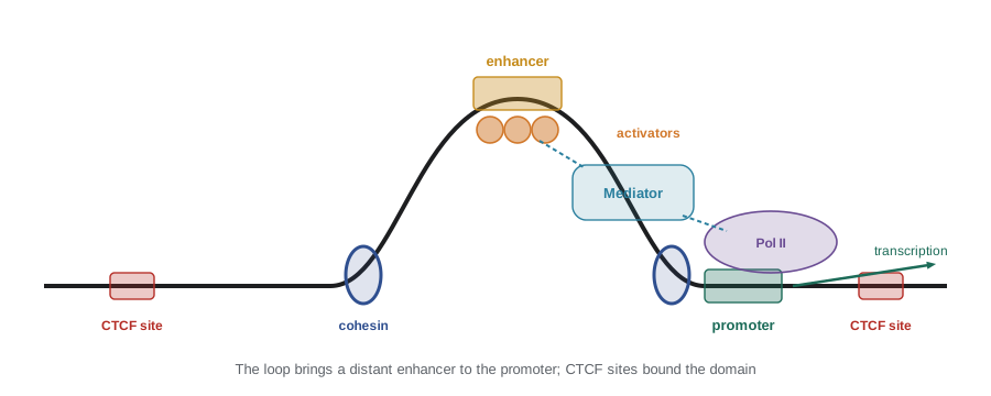
Enhancers reach their promoters by looping. Cohesin rings extrude DNA loops until they meet CTCF bound at specific sites, which act as boundaries, or insulators. As a result, enhancers usually act on promoters within the same topologically associating domain (lesson 2) and not on genes beyond its boundaries. When a boundary is deleted, an enhancer can inappropriately activate a neighboring gene. This has been shown to cause limb malformations in people and mice, when deletions or rearrangements near the gene EPHA4 allow limb enhancers to activate other genes.
Combinatorial control explains how a limited set of transcription factors can produce a vast variety of expression patterns. An enhancer may require several factors to be present together, so that a gene is expressed only in cells where all of them coincide. With 1,600 factors, even requiring combinations of just three gives hundreds of millions of possible combinations, far more than needed to distinguish every cell type.
Worked problem
Combinatorial possibilities
Suppose a set of 10 transcription factors is available, each either present or absent in a given cell type. How many distinct combinations are there? If an enhancer requires a specific set of 3 factors all to be present, in how many of the combinations is it active?
Show solution
- Each factor is present or absent: 2¹⁰ = 1,024 combinations.
- The enhancer is active when its 3 required factors are present; the other 7 can be anything: 2⁷ = 128 combinations.
- Fraction active = 128 / 1,024 = 1/8.
Answer: 1,024 combinations; the enhancer is active in 128 of them (one eighth). Adding a repressor that must be absent would halve that again, showing how each extra requirement narrows where a gene is expressed.
Signals and nuclear receptors
Transcription factors connect gene expression to the outside world. Steroid hormones such as cortisol and estrogen are lipid-soluble and cross the plasma membrane. Inside the cell, they bind members of the nuclear receptor family, zinc-finger transcription factors that then bind hormone response elements in DNA and activate or repress target genes. The glucocorticoid receptor, for example, sits in the cytoplasm bound to chaperones until cortisol binds; it then moves into the nucleus. Synthetic glucocorticoids such as dexamethasone are widely used anti-inflammatory drugs because the activated receptor represses genes for inflammatory proteins. Other signals act at the cell surface and are relayed by kinase cascades that phosphorylate transcription factors, changing their activity, location or stability (lesson 8).
RNA interference and microRNAs
In 1998 Andrew Fire and Craig Mello showed that double-stranded RNA injected into the nematode C. elegans silenced the matching gene far more powerfully than single-stranded antisense RNA. This process, RNA interference (RNAi), earned them the 2006 Nobel Prize. Double-stranded RNA is cut by the enzyme Dicer into fragments of about 21 to 23 nucleotides; one strand of each fragment is loaded into an Argonaute protein in the RNA-induced silencing complex (RISC), which then finds and cuts matching mRNAs.
Cells use the same machinery to make their own regulatory RNAs, the microRNAs (miRNAs). The first, lin-4, was found in C. elegans by Victor Ambros and colleagues in 1993, and Gary Ruvkun’s group showed that it pairs with the 3′ untranslated region of the lin-14 mRNA to repress it. Ambros and Ruvkun received the 2024 Nobel Prize in Physiology or Medicine. Humans have over a thousand miRNA genes. Unlike siRNAs, most animal miRNAs pair imperfectly with their targets: what matters most is the seed, nucleotides 2 to 8 of the miRNA, which must match a site in the target’s 3′ UTR. Binding leads to reduced translation and faster decay of the mRNA. Because a 7-nucleotide seed match occurs often, each miRNA can regulate hundreds of targets, and more than half of human genes are predicted to be miRNA targets.
COMP = str.maketrans("ACGU", "UGCA")

def seed_site(mirna):
    """Target site complementary to miRNA nucleotides 2-8, written 5'->3' in the mRNA."""
    seed = mirna[1:8]
    return seed.translate(COMP)[::-1]

let7 = "UGAGGUAGUAGGUUGUAUAGUU"            # human let-7a
site = seed_site(let7)
utr = ("AAUGCUACCUCAGAUUGCAACCUACCUCAAUUUGCUACCUCAAGGAUCUACCUCAUUU")
hits = [i for i in range(len(utr) - 6) if utr[i:i+7] == site]
print("let-7 seed (nt 2-8):", let7[1:8])
print("matching site in mRNA:", site)
print("site positions in this 3' UTR:", hits)let-7 seed (nt 2-8): GAGGUAG matching site in mRNA: CUACCUC site positions in this 3' UTR: [4, 21, 34, 47]
RNA interference has become a laboratory tool for switching off genes and, more recently, a class of medicines. Patisiran, approved in 2018 in the United States and Canada, uses siRNA in lipid nanoparticles to silence the liver gene for transthyretin in hereditary transthyretin amyloidosis, a disease in which misfolded transthyretin damages nerves and the heart. Later siRNA drugs, chemically modified and attached to a sugar (GalNAc) that targets liver cells, can be injected just twice a year, as with inclisiran, which lowers LDL cholesterol by silencing PCSK9.
Long non-coding RNAs and translational control
Long non-coding RNAs, more than 200 nucleotides long and not translated, number in the tens of thousands in humans. Most remain poorly understood, but some have clear roles. XIST, which coats one X chromosome in female mammals and recruits silencing complexes, is the best example (BIOL-201 lesson 7). Others act as scaffolds that bring chromatin regulators to particular genes, or as decoys that soak up miRNAs.
Translation itself can be regulated gene by gene. A classic example is iron. When iron is scarce, iron regulatory proteins bind iron-responsive elements, hairpins in the untranslated regions of certain mRNAs. Binding to the hairpin in the 5′ UTR of ferritin mRNA blocks translation of ferritin, the iron-storage protein, because the cell needs its iron, not stored iron. Binding to hairpins in the 3′ UTR of transferrin receptor mRNA protects it from decay, increasing iron uptake. When iron is plentiful, the proteins release both mRNAs and the responses reverse. A single regulatory protein thus coordinates opposite responses in two mRNAs through the position of its binding sites.
Common mistakes
MistakeEnhancers must be located immediately upstream of the promoter.
InsteadEnhancers can act from upstream, downstream or within introns, often tens to hundreds of kilobases away, by looping.
MistakeAll histone methylation represses transcription.
InsteadThe effect depends on the residue: H3K4me3 marks active promoters, while H3K9me3 and H3K27me3 mark silent chromatin.
MistakeDoubling a transcription factor’s concentration always doubles occupancy.
InsteadOccupancy saturates; near or above Kd, doubling concentration raises occupancy much less.
MistakeAnimal microRNAs must pair perfectly along their whole length with targets.
InsteadMost animal miRNAs need only a match in the seed region (nucleotides 2–8).
MistakeDNA methylation at a CpG island promoter activates the gene.
InsteadPromoter CpG island methylation typically silences the gene.
Summary and key terms
- Eukaryotic expression is controlled at chromatin, transcription, processing, export, stability, translation and protein levels.
- Remodelers, histone modifications (for example acetylation, H3K4me3, H3K27me3, H3K9me3) and CpG methylation regulate access to DNA.
- CpG islands mark most promoters; their methylation silences genes, and methylated CpG has been depleted from the genome by mutation.
- Transcription factors have separable DNA-binding and activation domains; occupancy follows θ = [TF]/(Kd + [TF]), steepened by cooperativity.
- Enhancers act at a distance by looping, within domains bounded by CTCF and cohesin; combinatorial control specifies cell-type expression.
- Nuclear receptors convert hormone signals into transcriptional responses.
- RNAi, microRNAs (seed matching), lncRNAs and RNA-binding proteins regulate expression after transcription.
- SWI/SNF complex
- An ATP-dependent chromatin remodeler that moves, ejects or restructures nucleosomes.
- histone acetyltransferase (HAT)
- An enzyme that acetylates histone lysines, generally opening chromatin.
- CpG island
- A CpG-rich region, often at a promoter, usually unmethylated when the gene is active.
- transcription factor
- A protein that binds specific DNA sequences and regulates transcription.
- dissociation constant (Kd)
- The concentration of a binding protein at which half of its sites are occupied.
- enhancer
- A DNA element that increases transcription of a gene, often from a distance.
- insulator
- A DNA element, often bound by CTCF, that blocks enhancer action across a boundary.
- Mediator
- A large co-activator complex linking transcription factors to RNA polymerase II.
- microRNA
- A small RNA of about 22 nucleotides that represses target mRNAs, mainly through seed pairing.
- RNA interference
- Gene silencing triggered by double-stranded RNA through Dicer and RISC.
Practice problems
Work each problem on paper first, then reveal the answer to check.
How does histone acetylation promote transcription?
Show answer
It neutralizes lysine positive charges, loosening DNA contact, and creates binding sites for bromodomain proteins.
Which histone mark is associated with Polycomb repression?
Show answer
H3K27me3.
Why is the human genome depleted of CpG dinucleotides?
Show answer
Methylated cytosine deaminates to thymine, converting CpG to TpG over evolutionary time.
A factor has Kd = 5 nM. What fraction of sites is occupied at 15 nM (no cooperativity)?
Show answer
15 / (5 + 15) = 0.75.
What concentration of a factor with Kd = 8 nM gives 50 percent occupancy?
Show answer
8 nM.
Why does cooperativity produce a switch-like response?
Show answer
Occupancy rises steeply over a narrow range of concentration, approximated by a Hill coefficient above 1.
How can an enhancer 200 kb away act on a promoter?
Show answer
DNA looping, mediated by cohesin and bounded by CTCF, brings the enhancer close to the promoter.
What happens to a glucocorticoid receptor when cortisol binds?
Show answer
It releases chaperones, enters the nucleus and binds response elements to regulate target genes.
Which nucleotides of a microRNA form the seed?
Show answer
Nucleotides 2 to 8.
How do iron regulatory proteins have opposite effects on ferritin and transferrin receptor mRNAs?
Show answer
Binding the 5′ UTR hairpin blocks ferritin translation; binding 3′ UTR hairpins stabilizes transferrin receptor mRNA.
Lesson quiz · 5 questions
Score 60% or more to complete this lesson.
Create a free account to save your progress, take quizzes and earn a certificate.