
BIOL-350 · Lesson 4 of 12 · 32 min
Excitable membranes and the action potential
Animals sense, decide and act quickly, and almost all of that speed depends on electrical signals carried by excitable cells: neurons, muscle fibres and some endocrine and sensory cells. An escaping squid jets away within a fraction of a second of seeing a predator; a cockroach turns from a puff of air in about 20 milliseconds. This lesson explains how membranes generate and propagate the action potential, the all-or-none electrical impulse that carries information over long distances. BIOL-214 introduced ion pumps, channels and the resting membrane potential, including the Nernst and Goldman equations. Here we build on that foundation to explain the action potential quantitatively, using the model that Alan Hodgkin and Andrew Huxley developed from their experiments on the squid giant axon, and to ask how animals have made conduction faster.
The resting potential revisited
Every animal cell has a voltage across its plasma membrane, inside negative relative to outside, typically −60 to −90 millivolts in neurons and muscle. Two ingredients produce it. First, the sodium–potassium pump keeps the concentrations of potassium high inside and sodium high outside. Second, the resting membrane is much more permeable to potassium than to sodium, because many potassium leak channels are open. Potassium ions diffuse out down their concentration gradient, leaving behind unbalanced negative charge, until the electrical force pulling them back balances the chemical force pushing them out. The voltage at which the two balance for a single ion is its equilibrium potential, given by the Nernst equation.
At 20 °C, RT/F is about 25.3 mV, so for a monovalent cation the Nernst potential is about 58 mV times the base-10 logarithm of the concentration ratio. When a membrane is permeable to several ions, the resting potential lies between their equilibrium potentials, weighted by the relative permeabilities, as described by the Goldman–Hodgkin–Katz equation. Because the resting membrane is mostly permeable to potassium, the resting potential lies close to the potassium equilibrium potential, but a small sodium permeability pulls it a few millivolts toward the sodium equilibrium potential.
import math

R, F = 8.314, 96485.0
def nernst(z, out, inn, T=293.15):
    return 1000 * R * T / (z * F) * math.log(out / inn)

def ghk(pk, pna, pcl, ko, ki, nao, nai, clo, cli, T=293.15):
    num = pk * ko + pna * nao + pcl * cli
    den = pk * ki + pna * nai + pcl * clo
    return 1000 * R * T / F * math.log(num / den)

# Squid giant axon, approximate concentrations (mM) in sea water
ko, ki, nao, nai, clo, cli = 20, 400, 440, 50, 560, 52
print(f"E_K  = {nernst(1, ko, ki):6.1f} mV")
print(f"E_Na = {nernst(1, nao, nai):6.1f} mV")
print(f"E_Cl = {nernst(-1, clo, cli):6.1f} mV")
for label_, pna in (("rest   (PK:PNa:PCl = 1:0.04:0.45)", 0.04), ("peak   (PK:PNa:PCl = 1:20:0.45)", 20.0)):
print(f"{label_}: Vm = {ghk(1.0, pna, 0.45, ko, ki, nao, nai, clo, cli):6.1f} mV")E_K = -75.7 mV E_Na = 54.9 mV E_Cl = -60.0 mV rest (PK:PNa:PCl = 1:0.04:0.45): Vm = -59.9 mV peak (PK:PNa:PCl = 1:20:0.45): Vm = 42.4 mV
With the classic permeability ratios at rest, the Goldman equation gives a resting potential of about −60 mV, close to the measured value for squid axons. If sodium permeability rises about 500-fold, as it does at the peak of an action potential, the membrane potential swings to about +40 mV, approaching the sodium equilibrium potential. The action potential is therefore, at heart, a brief, dramatic change in relative permeability, and the ions that cross the membrane to produce it are a tiny fraction of those present. A rough calculation shows that only about one potassium ion in a million inside a squid axon needs to cross the membrane to charge the membrane capacitance during one impulse. The concentration gradients barely change, and the pump restores them slowly in the background.
Hodgkin and Huxley and the squid giant axon
The squid giant axon, up to a millimetre in diameter, controls the mantle muscles used in jet propulsion. Its size let Hodgkin and Huxley, working at Plymouth in England in the late 1940s and early 1950s, thread a wire electrode inside it and use the voltage clamp, a feedback circuit developed by Kenneth Cole and George Marmont, to hold the membrane potential at chosen values while measuring the current that flowed. By replacing external sodium with an impermeant cation, they separated the current into a sodium component and a potassium component. Their analysis, published in 1952, earned them the 1963 Nobel Prize in Physiology or Medicine, which they shared with John Eccles.
The voltage clamp revealed three features of the membrane. A depolarizing step produced an early inward current carried by sodium, which rose within a millisecond and then declined even though the voltage was held constant, and a delayed outward current carried by potassium, which rose more slowly and was sustained. The sodium conductance therefore had two processes: activation, which opened it quickly on depolarization, and inactivation, which closed it more slowly. The potassium conductance had only delayed activation. Hodgkin and Huxley described each conductance with hypothetical gating particles, m and h for sodium and n for potassium, whose probabilities of being in the permissive position changed with voltage and time.
Each gating variable relaxes toward a voltage-dependent steady state with a voltage-dependent time constant. Decades later, cloning and structural biology showed that the gating particles correspond to real molecular parts. Voltage-gated sodium and potassium channels have four voltage-sensing domains, each containing a positively charged S4 helix that moves outward when the membrane depolarizes, pulling open the channel's gate. Fast inactivation of sodium channels is produced by a short intracellular loop, the inactivation gate, that swings in to block the pore. The fourth power in the potassium term matches the four subunits of the potassium channel, a satisfying confirmation of a model built from electrical measurements alone.
Simulating an action potential
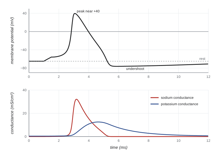
The Hodgkin–Huxley equations can be integrated numerically with a simple Euler method. The code below uses the standard parameters for the squid axon at 6.3 °C, written in the modern convention with a resting potential near −65 mV. It applies a brief current pulse and reports the main features of the resulting action potential, and then tests a weaker pulse to show the threshold.
import math

# Hodgkin-Huxley model (squid axon, 6.3 degC), modern sign convention
gNa, gK, gL = 120.0, 36.0, 0.3          # mS/cm^2
ENa, EK, EL = 50.0, -77.0, -54.4         # mV
Cm = 1.0                                 # uF/cm^2

def rates(V):
    am = 0.1 * (V + 40) / (1 - math.exp(-(V + 40) / 10))
    bm = 4.0 * math.exp(-(V + 65) / 18)
    ah = 0.07 * math.exp(-(V + 65) / 20)
    bh = 1.0 / (1 + math.exp(-(V + 35) / 10))
    an = 0.01 * (V + 55) / (1 - math.exp(-(V + 55) / 10))
    bn = 0.125 * math.exp(-(V + 65) / 80)
    return am, bm, ah, bh, an, bn

def run(amp, dur=0.5, T=20.0, dt=0.005):
    V = -65.0
    am, bm, ah, bh, an, bn = rates(V)
    m, h, n = am / (am + bm), ah / (ah + bh), an / (an + bn)
    peak, t_peak, vmin, t = V, 0.0, V, 0.0
    while t < T:
        I = amp if 1.0 <= t < 1.0 + dur else 0.0
        am, bm, ah, bh, an, bn = rates(V)
        m += dt * (am * (1 - m) - bm * m)
        h += dt * (ah * (1 - h) - bh * h)
        n += dt * (an * (1 - n) - bn * n)
        Iion = gNa * m**3 * h * (V - ENa) + gK * n**4 * (V - EK) + gL * (V - EL)
        V += dt * (I - Iion) / Cm
        if V > peak: peak, t_peak = V, t
        if t > t_peak and V < vmin: vmin = V
        t += dt
    return peak, t_peak, vmin

for amp in (5.0, 20.0, 80.0):
    peak, tp, vmin = run(amp)
    spike = "spike" if peak > 0 else "no spike"
    print(f"pulse {amp:4.1f} uA/cm2 x 0.5 ms: peak {peak:6.1f} mV at {tp:4.2f} ms, "
f"after-hyperpolarization {vmin:6.1f} mV ({spike})")pulse 5.0 uA/cm2 x 0.5 ms: peak -62.8 mV at 1.50 ms, after-hyperpolarization -65.6 mV (no spike) pulse 20.0 uA/cm2 x 0.5 ms: peak 39.6 mV at 3.06 ms, after-hyperpolarization -76.2 mV (spike) pulse 80.0 uA/cm2 x 0.5 ms: peak 41.8 mV at 1.82 ms, after-hyperpolarization -76.2 mV (spike)
The weakest pulse produces only a small, passive depolarization that decays; the stronger pulses produce a full action potential peaking near +40 mV, followed by an undershoot below the resting potential. A pulse four times stronger produces a spike of essentially the same size; it simply arrives sooner, because threshold is reached faster. This is the all-or-none property: once threshold is crossed, the positive feedback between depolarization and sodium channel opening runs to completion, and the shape of the impulse is set by the membrane, not by the stimulus. Information is therefore carried not by the size of action potentials but by their frequency and timing.
Threshold, refractoriness and firing patterns
Threshold is the voltage at which the inward sodium current produced by a depolarization just exceeds the outward potassium and leak currents, so that the depolarization becomes self-sustaining. After an action potential, sodium channels remain inactivated for a millisecond or so, and potassium channels remain open, so a second impulse cannot be triggered at all during the absolute refractory period and needs a stronger stimulus during the relative refractory period. Refractoriness limits the maximum firing frequency, to several hundred impulses per second in fast neurons, and it makes action potentials travel in one direction along an axon, because the membrane behind an advancing impulse is refractory.
Real neurons contain many more kinds of channels than the squid axon. Calcium channels, calcium-activated potassium channels, hyperpolarization-activated cation channels and slowly inactivating potassium channels give different neurons different firing patterns: regular firing, adaptation in which the rate slows during a sustained stimulus, bursting, and rhythmic pacemaking. The heart's pacemaker cells and the neurons that drive breathing and walking use these channels to generate rhythms without any outside input. Comparative studies show that the basic voltage-gated sodium and potassium channels are ancient, with homologues in single-celled organisms, while the diversity of channel types has expanded greatly in animals with complex nervous systems.
Toxins that target channels
Many animals and plants defend themselves with toxins that block or modify ion channels, and these toxins have been invaluable tools. Tetrodotoxin, made by bacteria living in pufferfish, rough-skinned newts of the Pacific Northwest and blue-ringed octopuses, blocks voltage-gated sodium channels from the outside. Saxitoxin, made by dinoflagellates and concentrated by shellfish during harmful algal blooms, does the same and causes paralytic shellfish poisoning, which is why shellfish harvesting on both coasts is closed when monitoring detects it. Tetraethylammonium blocks potassium channels. Garter snakes that feed on rough-skinned newts have evolved sodium channels resistant to tetrodotoxin, and the newts in turn have evolved higher toxin levels, a famous example of a coevolutionary arms race studied by Edmund Brodie and colleagues.
Passive spread and the cable properties of axons
An action potential spreads along an axon because the current that enters at an active patch of membrane flows along the axon's interior and depolarizes the membrane ahead to threshold. How far this local current spreads depends on the axon's cable properties. Current flowing along the inside of the axon meets the axial resistance of the cytoplasm, while some of it leaks out across the membrane through the membrane resistance. The length constant, λ, is the distance over which a steady voltage decays to about 37 per cent of its starting value. It increases with the ratio of membrane resistance to internal resistance, and because the internal resistance per unit length falls with the square of the diameter while the membrane resistance per unit length falls only with the diameter, λ increases with the square root of axon diameter.
Membrane capacitance adds a time element. Before the membrane voltage can change, its capacitance must be charged, and the time constant τ, the product of membrane resistance and capacitance, describes how quickly this happens. Conduction velocity in an unmyelinated axon depends on both: a larger λ means current reaches farther ahead, and a smaller τ means the membrane there charges faster. Theory and measurements agree that conduction velocity in unmyelinated axons is roughly proportional to the square root of diameter. That explains the giant axons of squid, earthworms and cockroaches: doubling conduction velocity requires a fourfold increase in diameter, which is affordable only for a few axons dedicated to escape.
Myelin and saltatory conduction
Vertebrates found a better solution. In the peripheral nervous system, Schwann cells, and in the central nervous system, oligodendrocytes, wrap axons in many layers of membrane called myelin. Myelin increases the membrane resistance and decreases the capacitance of the wrapped segments, so current spreads much farther with little loss and charges the membrane much less. Voltage-gated sodium channels are concentrated at gaps in the myelin, the nodes of Ranvier, about 1 to 2 µm long and spaced 0.2 to 2 mm apart. The action potential appears to jump from node to node, saltatory conduction, and conduction velocity becomes roughly proportional to diameter rather than to its square root. A myelinated axon 10 µm across conducts at about 50 to 60 metres per second, about the same speed as a squid giant axon 50 to 100 times wider.
| Axon | Diameter | Myelinated? | Conduction velocity (approx.) | Function |
|---|---|---|---|---|
| Squid giant axon | 0.5–1 mm | No | 20–25 m/s | Escape jet |
| Earthworm medial giant fibre | about 0.05–0.1 mm | Partly wrapped (loose sheath) | up to about 30 m/s | Withdrawal into burrow |
| Cockroach giant interneuron | about 0.05 mm | No | about 7–10 m/s | Escape turn |
| Mammalian C fibre (pain, temperature) | 0.2–1.5 µm | No | 0.5–2 m/s | Slow pain, warmth |
| Mammalian Aα motor axon | 12–20 µm | Yes | 70–120 m/s | Skeletal muscle control |
| Shrimp (Penaeus) giant fibre | about 0.1 mm | Yes (unusual myelin) | over 90 m/s | Tail-flip escape |
Myelin evolved in jawed vertebrates and, independently, in some invertebrates, notably certain shrimp and copepods, whose myelin-like sheaths support very fast conduction in small axons. Myelination has a cost: myelin must be made and maintained, and in demyelinating diseases such as multiple sclerosis, loss of myelin slows or blocks conduction. But it allows vertebrates to pack many fast axons into a small nerve, which is essential for complex, rapid behaviour in large bodies. A human sciatic nerve contains tens of thousands of axons; if they all had to be unmyelinated and as fast as they are, the nerve would be many centimetres thick.
import math

# Conduction velocity scaling (rules of thumb):
#   unmyelinated: v ~ k1 * sqrt(d)      myelinated: v ~ k2 * d  (d in um)
k1 = 22.0 / math.sqrt(500.0)   # calibrated: squid axon, 500 um -> 22 m/s
k2 = 5.5                       # about 5.5 m/s per um (Hursh-type rule for mammals)

print(" target v (m/s)   unmyelinated d needed   myelinated d needed")
for v in (2, 10, 25, 60, 100):
    d_un = (v / k1) ** 2
    d_my = v / k2
    print(f"   {v:6.0f}         {d_un:10.0f} um            {d_my:6.1f} um")

# time for a signal from a moose's brain to a hind foot about 2.5 m away
for v in (1.0, 60.0):
print(f"2.5 m at {v:5.1f} m/s takes {2.5 / v * 1000:7.0f} ms") target v (m/s) unmyelinated d needed myelinated d needed
2 4 um 0.4 um
10 103 um 1.8 um
25 646 um 4.5 um
60 3719 um 10.9 um
100 10331 um 18.2 um
2.5 m at 1.0 m/s takes 2500 ms
2.5 m at 60.0 m/s takes 42 msTo reach 60 metres per second without myelin, an axon would need to be almost 4 mm across; with myelin, about 11 µm suffices. For a large animal, conduction delays matter. A signal from a moose's brain to its hind foot would take more than two seconds at the speed of a slow unmyelinated fibre, but about 40 milliseconds in a fast myelinated motor axon. Even so, large animals live with longer neural delays than small ones, and studies of reaction times across mammals show that the largest species respond more slowly to disturbances in proportion to their size.
Worked problem
Interpreting a voltage-clamp experiment
A squid axon is voltage-clamped and stepped from −65 mV to 0 mV. The total current shows a brief inward phase followed by a sustained outward phase. When external sodium is replaced by choline, the inward phase disappears and the outward phase is unchanged. When tetraethylammonium is added instead, the outward phase disappears and the inward phase now rises and then decays back to near zero. What can you conclude?
Show solution
- Removing external sodium abolishes the early inward current, so it is carried by sodium ions moving in.
- Tetraethylammonium, a potassium channel blocker, abolishes the late outward current, so that current is carried by potassium moving out.
- With potassium current blocked, the sodium current still turns off during the maintained step, so sodium channels inactivate even when the voltage stays depolarized.
- The potassium current is sustained during the step, so delayed rectifier potassium channels activate slowly and do not inactivate on this time scale.
Answer: The membrane has a fast-activating, inactivating sodium conductance and a slower, non-inactivating potassium conductance. These are exactly the two components Hodgkin and Huxley used to build their model of the action potential.
Common mistakes
MistakeThinking that large amounts of sodium and potassium cross the membrane in each action potential.
InsteadOnly a tiny fraction of the ions present cross; concentration gradients barely change and the pump restores them slowly.
MistakeSaying the falling phase of the action potential is caused by the sodium pump.
InsteadIt is caused by sodium channel inactivation and the opening of voltage-gated potassium channels.
MistakeAssuming stronger stimuli produce bigger action potentials.
InsteadAction potentials are all-or-none; stronger stimuli raise firing frequency, not spike size.
MistakeStating that myelin speeds conduction by making the action potential travel faster through the myelin.
InsteadMyelin raises membrane resistance and lowers capacitance so passive current spreads farther and faster between nodes, where spikes regenerate.
MistakeExpecting conduction velocity in unmyelinated axons to be proportional to diameter.
InsteadIt is roughly proportional to the square root of diameter; in myelinated axons it is roughly proportional to diameter.
MistakeTreating the resting potential as equal to the potassium equilibrium potential.
InsteadSmall sodium and chloride permeabilities pull it a few millivolts away, as the Goldman equation shows.
Summary and key terms
- The resting potential arises from ion gradients maintained by the Na⁺/K⁺ pump and a membrane mostly permeable to K⁺.
- The Nernst equation gives each ion's equilibrium potential; the Goldman equation combines ions by permeability.
- Voltage-clamp experiments on the squid giant axon revealed fast-activating, inactivating Na⁺ currents and delayed, sustained K⁺ currents.
- The Hodgkin–Huxley model reproduces the action potential from gating variables that correspond to real channel structures.
- Action potentials are all-or-none; refractoriness limits firing frequency and enforces one-way propagation.
- Conduction velocity depends on cable properties; it scales with √d in unmyelinated axons and with d in myelinated axons.
- Myelin and saltatory conduction let vertebrates pack many fast, small axons into nerves; giant axons are the invertebrate solution.
- equilibrium potential
- The membrane potential at which the electrical and chemical forces on an ion balance.
- Goldman–Hodgkin–Katz equation
- An equation giving the membrane potential from ion concentrations and relative permeabilities.
- voltage clamp
- A technique that holds membrane voltage constant while measuring the current that flows.
- activation
- The voltage-dependent opening of a channel's gate.
- inactivation
- Closure of a channel by a separate gate during maintained depolarization.
- threshold
- The membrane potential at which depolarization becomes self-sustaining and an action potential fires.
- refractory period
- The interval after an action potential during which another is impossible or harder to trigger.
- length constant
- The distance over which a steady voltage along an axon decays to about 37 per cent.
- membrane time constant
- The product of membrane resistance and capacitance, setting how fast the membrane voltage changes.
- myelin
- Layers of glial membrane wrapped around an axon that increase resistance and reduce capacitance.
- node of Ranvier
- A gap in the myelin where voltage-gated sodium channels are concentrated.
- saltatory conduction
- Propagation of action potentials from node to node along a myelinated axon.
Practice problems
Work each problem on paper first, then reveal the answer to check.
At 20 °C, calculate E_K for [K⁺]o = 5 mM and [K⁺]i = 150 mM.
Show answer
58 × log(5/150) ≈ 58 × (−1.48) ≈ −86 mV.
Why is the resting potential usually a few millivolts less negative than E_K?
Show answer
A small resting permeability to Na⁺ (and Cl⁻ effects) pulls it toward E_Na, as the Goldman equation predicts.
What two processes control sodium conductance in the Hodgkin–Huxley model?
Show answer
Activation (m gates) and inactivation (h gate).
What is the effect of tetrodotoxin on an axon, and why?
Show answer
It blocks voltage-gated sodium channels, so action potentials cannot be generated.
Explain why action potentials travel in only one direction along an axon.
Show answer
The membrane behind the impulse is refractory because its sodium channels are inactivated.
How does conduction velocity change if an unmyelinated axon's diameter quadruples?
Show answer
It roughly doubles (√4 = 2).
Name the glial cells that form myelin in the peripheral and central nervous systems.
Show answer
Schwann cells (peripheral) and oligodendrocytes (central).
Why do squid have giant axons rather than myelin?
Show answer
Cephalopods lack vertebrate-type myelin; a few very wide axons provide fast conduction for escape.
What is the length constant, and how does it depend on axon diameter?
Show answer
The distance over which voltage decays to 1/e; it increases with the square root of diameter.
How do garter snakes resist tetrodotoxin from newts?
Show answer
Mutations in their sodium channels reduce toxin binding.
Lesson quiz · 5 questions
Score 60% or more to complete this lesson.
Create a free account to save your progress, take quizzes and earn a certificate.