
BIOL-107 · Lesson 6 of 12 · 26 min
Membranes: structure and fluidity
Every cell is wrapped in a membrane only about 7 to 10 nanometres thick, thinner than a soap bubble's film and roughly ten thousand times thinner than a sheet of paper. That film separates life from non-life: inside, the cell keeps its own chemistry; outside is the environment. Eukaryotic cells also use membranes to build their internal compartments. This lesson explains what membranes are made of, why they form spontaneously, why they are fluid, and how cells adjust that fluidity. Lesson 7 then shows how things cross them.
A brief history of the membrane model
Scientists inferred the structure of the membrane long before they could see it, by a chain of clever experiments. In 1925 Evert Gorter and François Grendel extracted the lipids from red blood cell membranes, spread them as a single layer on water, and found the area was about twice the surface area of the cells. They concluded that the membrane is a lipid bilayer. (Their measurements contained two errors that happened to cancel out, but the conclusion was right.) Because pure lipid bilayers did not fully explain how membranes behave, models with protein layers coating the lipid were proposed, but by the late 1960s electron microscopy and biochemistry showed that many proteins pass right through the bilayer.
In 1972 S. J. Singer and Garth Nicolson proposed the fluid mosaic model, which is still the basis of how we picture membranes: a fluid bilayer of lipids in which proteins float like icebergs, forming a mosaic. A famous experiment by Larry Frye and Michael Edidin in 1970 had already shown the fluidity. They fused a mouse cell with a human cell, each with its surface proteins labelled with a different colour of fluorescent antibody. At first the colours stayed on their own halves of the hybrid cell, but within about 40 minutes at 37 °C they had completely intermixed. At low temperature, mixing slowed dramatically. Membrane proteins drift sideways in a fluid layer.
Phospholipids and the bilayer
The main structural molecules of membranes are phospholipids (lesson 3). Each has a hydrophilic head, containing a negatively charged phosphate group and usually a small attached molecule such as choline, and two hydrophobic fatty acid tails. In water, phospholipids spontaneously arrange themselves so their tails avoid water: they form a bilayer, two layers with the tails facing each other in the middle and the heads facing the water on both sides. No energy or enzyme is needed; the hydrophobic effect drives it. A bilayer also tends to close on itself into a sealed sphere, because open edges would expose tails to water. That is why membranes form closed compartments.
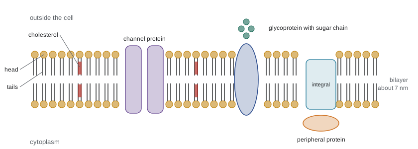
The hydrophobic core, about 3 to 4 nm thick, is what makes a membrane a barrier. Small non-polar molecules such as oxygen, carbon dioxide and steroid hormones dissolve in it and pass through easily. Water passes slowly. Ions and large polar molecules such as glucose barely cross a pure bilayer at all; they need protein help (lesson 7).
# Relative permeability of a pure lipid bilayer (approximate, cm/s, log scale)
import math
P = {"oxygen O2": 2e1, "carbon dioxide CO2": 3e0, "water H2O": 3e-3,
     "urea": 4e-6, "glycerol": 5e-6, "glucose": 4e-10,
     "chloride Cl-": 1e-11, "potassium K+": 5e-14, "sodium Na+": 1e-14}
for name, p in P.items():
    bar = "#" * int(math.log10(p) + 15)
print(f"{name:<20} {p:8.0e} {bar}")oxygen O2 2e+01 ################ carbon dioxide CO2 3e+00 ############### water H2O 3e-03 ############ urea 4e-06 ######### glycerol 5e-06 ######### glucose 4e-10 ##### chloride Cl- 1e-11 #### potassium K+ 5e-14 # sodium Na+ 1e-14 #
The output spans more than 15 powers of ten. Oxygen crosses a bilayer roughly a million billion (10 to the power 15) times more easily than a sodium ion (the values are approximate and vary with lipid composition). The permeability gap is the whole point: by blocking ions and most polar molecules, the membrane lets the cell build up very different concentrations on each side and then control traffic with proteins.
The other membrane lipids
Membranes contain many kinds of phospholipids, differing in their heads and tails, and two other important classes of lipid:
| Lipid | Structure | Role |
|---|---|---|
| Phosphatidylcholine | Phospholipid with a choline head | The most common phospholipid in animal membranes, mostly in the outer layer |
| Phosphatidylserine | Phospholipid with a negatively charged serine head | Kept in the inner layer; appears on the outer surface when a cell dies, signalling “eat me” to scavenger cells |
| Phosphatidylinositol | Phospholipid with an inositol sugar head | Source of signalling molecules inside the cell (lesson 11) |
| Cholesterol | Steroid with a small hydroxyl head | Up to about 30 to 40 per cent of lipid molecules in some animal plasma membranes; buffers fluidity |
| Glycolipids | Lipids with sugar chains attached | Outer surface only; cell recognition and blood group antigens |
The two layers of a membrane are not identical. Cells actively maintain different lipid compositions in the inner and outer layers, a property called asymmetry. Lipids drift freely sideways, exchanging places with neighbours millions of times a second, but they rarely flip from one layer to the other on their own, because the polar head would have to cross the hydrophobic core. Enzymes called flippases and scramblases move them when needed.
Membrane fluidity and how cells control it
At body temperature a membrane behaves like a two-dimensional liquid with the consistency of light oil. Fluidity matters: membrane proteins must move and change shape to work, vesicles must fuse, and cells must divide. If a membrane is too fluid it leaks; if too stiff it cracks and its proteins stop working. Three factors set the fluidity:
| Factor | Effect on fluidity | Reason |
|---|---|---|
| Temperature | Higher temperature increases fluidity | Molecules move faster; below a transition temperature the bilayer becomes a stiff gel |
| Unsaturated fatty acid tails | Increase fluidity | Cis double bonds kink the tails so they cannot pack tightly |
| Tail length | Shorter tails increase fluidity | Fewer van der Waals contacts between neighbouring tails |
| Cholesterol | Buffers fluidity: reduces it at high temperature, prevents stiffening at low temperature | Its rigid rings restrain tail movement but also stop tails packing into a gel |
Organisms that cannot control their body temperature adjust their membranes instead, a process called homeoviscous adaptation. Bacteria grown in the cold make more unsaturated and shorter fatty acids; grown warm, they make more saturated ones. Fish in Arctic waters have more unsaturated membrane lipids than tropical fish. Winter wheat and other cold-hardy plants increase membrane unsaturation in autumn. Reindeer even vary the fatty acid composition of cell membranes along their legs, with more unsaturated lipids near the cold hooves.
Worked problem
Predicting membrane changes
A culture of bacteria growing at 37 °C is moved to 15 °C. Predict how the composition of its membrane lipids will change over the next few hours, and explain why.
Show solution
- Lower temperature slows lipid movement, so the membrane becomes more viscous and may approach its gel transition.
- To restore normal fluidity, the cell needs lipids that pack less tightly.
- It increases the proportion of unsaturated fatty acids (cis double bonds create kinks) and of shorter fatty acid chains.
- Enzymes called desaturases, which insert double bonds, become more active.
Answer: The bacteria will make more unsaturated and shorter-chain fatty acids, keeping their membranes fluid at the lower temperature.
Membrane proteins
If lipids form the fabric of the membrane, proteins do most of the work. By mass, membranes are typically about half protein, though this varies from about 25 per cent in the myelin sheath around nerve fibres to about 75 per cent in the inner mitochondrial membrane, which is packed with the machinery of respiration. Proteins are classified by how they associate with the bilayer.
Integral proteins are embedded in the membrane. Most span it completely (transmembrane proteins), with hydrophobic stretches of about 20 amino acids that coil into alpha helices across the core and hydrophilic parts sticking out on each side. Some cross once; others, such as transporters and channels, weave back and forth many times. Peripheral proteins sit on one surface, attached to integral proteins or to lipid heads, and can be removed with gentle treatments such as salt solutions.
| Function | Example |
|---|---|
| Transport | Channels for ions, carriers for glucose, pumps such as the sodium–potassium pump |
| Enzymatic activity | Enzymes of the respiratory chain in the inner mitochondrial membrane |
| Signal reception | Receptors for hormones such as insulin |
| Cell recognition | Glycoproteins that identify cells to the immune system (MHC proteins) |
| Intercellular joining | Proteins of tight junctions and desmosomes |
| Attachment to the cytoskeleton and matrix | Integrins linking actin inside to collagen outside |
# How many amino acids does a membrane-spanning alpha helix need?
core_thickness_nm = 3.0      # hydrophobic core
rise_per_residue_nm = 0.15   # an alpha helix rises 0.15 nm per amino acid
print("Residues to span the core:", round(core_thickness_nm / rise_per_residue_nm))

# A protein that crosses the membrane 12 times (like many transporters)
crossings = 12
print("Minimum hydrophobic residues in transmembrane helices:", crossings * 20)Residues to span the core: 20 Minimum hydrophobic residues in transmembrane helices: 240
Programs that predict membrane proteins from a gene sequence look for exactly these stretches of about 20 hydrophobic amino acids. Roughly a quarter to a third of human genes encode membrane proteins, and more than half of all medicines act on membrane proteins, particularly receptors, channels and transporters.
Proteins also move in the membrane, but many are not free to wander. They may be tethered to the cytoskeleton underneath, held by junctions to neighbouring cells, or gathered in particular regions. Cells lining the gut, for example, keep glucose transporters on the side facing the gut and different transporters on the side facing the blood, separated by tight junctions. Small, more ordered patches enriched in cholesterol and certain lipids, often called lipid rafts, may help cluster some proteins, though their size and nature are still debated.
The carbohydrate coat
On the outer surface of animal cells, short chains of sugars attached to proteins and lipids form a fuzzy layer called the glycocalyx. These sugar chains are like name tags: they help cells recognize each other, let sperm recognize eggs, guide white blood cells to sites of infection, and define blood groups. The A, B and O blood types depend on which sugar, if any, is added to the end of a chain on red blood cells. A blood transfusion with the wrong type triggers an immune attack on the foreign sugars.
Membranes across life
All cell membranes follow the same general plan, with interesting variations. Archaea, many of which live in hot or acidic places, have distinctive lipids with ether rather than ester links and branched tails; some even form a single layer whose molecules span the whole membrane, resisting heat. Plant cells use plant sterols in place of cholesterol. The inner mitochondrial membrane contains a special phospholipid, cardiolipin, shared with bacteria, another clue to its bacterial origin (lesson 1).
Membrane biology matters medically. Some antibiotics, such as polymyxins, disrupt bacterial membranes. Anesthetic drugs and alcohol interact with membranes and membrane proteins. Many viruses, including influenza and SARS-CoV-2, are wrapped in a membrane taken from their host cell and enter new cells by fusing their membrane with the cell's. Soap destroys such viruses by dissolving their membranes, which is why hand washing works so well.
Common mistakes
MistakeThe cell membrane is a rigid wall.
InsteadIt is a fluid bilayer in which lipids and many proteins drift sideways; the rigid layer in plants and bacteria is the separate cell wall.
MistakeCholesterol always makes membranes less fluid.
InsteadCholesterol buffers fluidity: it reduces fluidity at high temperature but prevents the membrane stiffening into a gel at low temperature.
MistakeLipids flip freely between the two layers of the membrane.
InsteadLipids move sideways rapidly but rarely flip across without enzymes, so the two layers can differ in composition.
MistakeIons cross membranes easily because they are small.
InsteadTheir charge and shell of water molecules keep ions out of the hydrophobic core; they need channel or carrier proteins.
MistakeAll membrane proteins span the membrane.
InsteadPeripheral proteins sit on one surface without crossing the core.
Summary and key terms
- Membranes are 7 to 10 nm thick and form spontaneously as phospholipid bilayers because of the hydrophobic effect.
- The fluid mosaic model describes proteins floating in a fluid lipid bilayer; the Frye–Edidin cell fusion experiment showed proteins move sideways.
- The hydrophobic core lets gases and small non-polar molecules through but blocks ions and most polar molecules.
- Fluidity rises with temperature, unsaturation and shorter tails; cholesterol buffers fluidity in both directions.
- Organisms adjust membrane lipids to keep fluidity steady at different temperatures (homeoviscous adaptation).
- Integral proteins span or embed in the bilayer with hydrophobic helices of about 20 amino acids; peripheral proteins sit on the surface.
- Membrane proteins transport substances, act as enzymes, receive signals, recognize cells and anchor structures; sugar chains on the outer surface form the glycocalyx.
- lipid bilayer
- A double layer of phospholipids with hydrophobic tails inside and hydrophilic heads facing water.
- fluid mosaic model
- The model of membranes as a fluid lipid bilayer with embedded and attached proteins.
- membrane fluidity
- How freely lipids and proteins move within a membrane.
- cholesterol
- A steroid in animal membranes that buffers fluidity.
- integral protein
- A protein embedded in the membrane, often spanning it.
- transmembrane protein
- An integral protein that crosses the entire bilayer.
- peripheral protein
- A protein attached to the membrane surface without entering the hydrophobic core.
- glycocalyx
- The carbohydrate-rich layer on the outer surface of animal cells.
- membrane asymmetry
- The difference in lipid and protein composition between the two layers of a membrane.
- homeoviscous adaptation
- Adjustment of membrane lipid composition to keep fluidity constant as temperature changes.
Practice problems
Work each problem on paper first, then reveal the answer to check.
Why do phospholipids form bilayers spontaneously in water?
Show answer
Their hydrophobic tails avoid water and cluster together while the hydrophilic heads face water; this arrangement is favoured by the hydrophobic effect.
Describe the Frye–Edidin experiment and its conclusion.
Show answer
Mouse and human cells with differently labelled surface proteins were fused; the labels mixed within about 40 minutes at 37 °C, showing membrane proteins move sideways in a fluid bilayer.
Rank oxygen, glucose and potassium ions by how easily they cross a pure lipid bilayer.
Show answer
Oxygen (easily) > glucose (very slowly) > potassium ions (almost not at all).
How does cholesterol affect membrane fluidity at high and low temperatures?
Show answer
At high temperature it restrains tails and reduces fluidity; at low temperature it prevents tight packing and keeps the membrane fluid.
Predict how the membranes of a fish change as it moves from warm to cold water.
Show answer
It increases the proportion of unsaturated (and shorter) fatty acids to keep the membranes fluid.
About how many amino acids are needed for an alpha helix to span the hydrophobic core?
Show answer
About 20, since the core is about 3 nm and each residue rises 0.15 nm.
Why does the inner mitochondrial membrane have such a high protein content?
Show answer
It holds the protein complexes of the electron transport chain and ATP synthase.
Give four functions of membrane proteins with examples.
Show answer
Transport (sodium–potassium pump), enzyme (respiratory chain), receptor (insulin receptor), recognition (MHC glycoproteins); also joining and anchoring.
What is the glycocalyx, and what is one of its functions?
Show answer
A layer of sugar chains on glycoproteins and glycolipids outside the cell; it helps cells recognize each other, as in blood groups.
Why does washing with soap inactivate viruses such as influenza?
Show answer
Soap disrupts the lipid membrane envelope that surrounds these viruses.
Lesson quiz · 5 questions
Score 60% or more to complete this lesson.
Create a free account to save your progress, take quizzes and earn a certificate.