
BIOL-360 · Lesson 8 of 12 · 28 min
Neural development and the neural crest
The vertebrate nervous system is the most complex structure produced by any embryo. A human brain contains roughly 86 billion neurons, each connected to thousands of others, and all of it develops from a flat sheet of ectoderm a few millimeters long. This lesson follows the early steps: the formation of the neural tube, its subdivision along the anterior–posterior and dorsal–ventral axes, the generation of neurons from progenitor cells, the remarkable migratory cells of the neural crest, and the guidance of axons to their targets. The physiology of neurons and synapses was covered in BIOL-350 lessons 4 and 5, and BIOL-420 takes up synaptic plasticity, sensory and motor systems and memory; here the focus is on how the nervous system is built.
From neural plate to neural tube
Lesson 5 showed how the organizer induces the dorsal ectoderm to become neural plate by blocking BMP. The neural plate then rolls up into the neural tube in a process called neurulation. In primary neurulation, used for the brain and most of the spinal cord, the edges of the plate thicken and rise as neural folds, the plate bends at hinge points, and the folds meet at the dorsal midline and fuse, separating the neural tube from the overlying epidermis. Cell shape changes drive bending: cells at the medial hinge point become wedge-shaped by apical constriction, and convergent extension narrows and lengthens the plate. In secondary neurulation, used for the most posterior part of the spinal cord, a solid cord of cells forms and then hollows out.
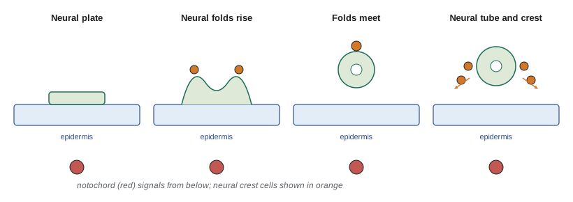
In humans, neurulation occurs between about 22 and 28 days after fertilization, often before a woman knows she is pregnant. Closure begins in the region of the future neck and proceeds in both directions, with the anterior neuropore closing around day 25 and the posterior neuropore around day 27 or 28. Failure of closure causes neural tube defects. Failure at the anterior end causes anencephaly, in which the forebrain does not develop, and is fatal. Failure at the posterior end causes spina bifida, in which the spinal cord remains open or covered only by a membrane, with effects ranging from mild to paralysis of the legs and loss of bladder control.
Neural tube defects have genetic and environmental causes. Mutations in genes of the planar cell polarity pathway, which drives convergent extension, cause severe defects in mice, and variants in these genes have been found in human patients. Maternal diabetes, obesity, high fever and the anticonvulsant valproic acid increase risk. The most important environmental factor is maternal folate. A landmark trial by the UK Medical Research Council, published in 1991, showed that folic acid supplements reduced recurrence of neural tube defects by about 70 percent. Both Canada and the United States made fortification of white flour and some other grain products with folic acid mandatory in 1998.
In the United States, studies after fortification found that the prevalence of neural tube defects fell by roughly a quarter to a third, and the Centers for Disease Control and Prevention estimate that fortification prevents over a thousand affected pregnancies each year. The US Preventive Services Task Force recommends that all people who could become pregnant take a daily supplement containing 0.4 to 0.8 mg of folic acid. Since 2016, the Food and Drug Administration has permitted voluntary fortification of corn masa flour, which matters for communities that eat more corn than wheat products.
Why folate prevents neural tube defects is not fully understood. Folate is needed to make nucleotides for the rapid cell division of the neural plate and for the methylation reactions that regulate gene expression. Mice with mutations in folate metabolism enzymes show neural tube defects, and some forms of defect resistant to folic acid respond instead to inositol in animal studies.
Patterning the neural tube
The neural tube is patterned along both axes. Along the anterior–posterior axis, it bulges into three primary brain vesicles, the forebrain (prosencephalon), midbrain (mesencephalon) and hindbrain (rhombencephalon), followed by the spinal cord. The forebrain and hindbrain later subdivide further. The hindbrain is divided into seven or eight segments called rhombomeres, which are lineage-restricted compartments; cells do not mix between them. Hox genes of paralogous groups 1 to 4 are expressed in a nested pattern in the rhombomeres and specify their identity, under the control of retinoic acid from the adjacent mesoderm. Local signaling centers refine the pattern: the isthmic organizer at the midbrain–hindbrain boundary secretes FGF8 and Wnt1 and patterns the midbrain and cerebellum, and grafting it to the forebrain induces midbrain-like structures. Along the dorsal–ventral axis, Sonic hedgehog from the notochord and floor plate, and BMPs and Wnts from the roof plate, pattern progenitor domains, as described in lesson 6. Ventral progenitors give rise to motor neurons and some interneurons; dorsal progenitors give rise to sensory relay neurons.
Making neurons
The wall of the neural tube begins as a single layer of neuroepithelial cells, whose nuclei move up and down as they progress through the cell cycle, a process called interkinetic nuclear migration: they divide at the apical surface facing the lumen and replicate their DNA near the basal surface. These cells become radial glia, long cells spanning the wall of the tube, which act both as stem cells and as scaffolds along which young neurons migrate. Radial glial cells can divide symmetrically to make more progenitors or asymmetrically to make one progenitor and one neuron or intermediate progenitor. Notch signaling, through lateral inhibition, keeps a pool of progenitors while some cells differentiate; proneural transcription factors of the basic helix-loop-helix family, such as Neurogenin and Ascl1, drive differentiation.
In the mammalian cerebral cortex, neurons are produced in an inside-out sequence. The first neurons to be born form the deepest layer, and successive waves migrate past them to form progressively more superficial layers, so the six-layered cortex is built from the inside out. Disruption of migration causes malformations such as lissencephaly, a smooth brain lacking normal folds; mutations in genes for the extracellular protein reelin or the microtubule regulator LIS1 cause such defects. The human cortex is expanded relative to other primates partly because of an expanded outer subventricular zone containing outer radial glia, which provide many more rounds of neuron production. Some human-specific genes, such as NOTCH2NL, arose by recent gene duplication and promote progenitor proliferation.
The neural crest
The neural crest arises at the border between neural plate and epidermis. As the neural folds close, crest cells undergo an epithelial–mesenchymal transition, delaminate from the dorsal neural tube and migrate throughout the embryo along defined pathways, forming an astonishing range of derivatives. Because of this multipotency and its importance in vertebrate evolution, the neural crest is sometimes called the fourth germ layer. Crest cells are specified by intermediate BMP levels together with Wnt and FGF signals at the neural plate border, which activate border specifier genes such as Pax3 and Msx1, followed by neural crest specifiers such as Snail2, FoxD3 and Sox10.
| Region of origin | Main derivatives |
|---|---|
| Cranial (head) | Cartilage, bone and connective tissue of the face and jaws, cranial nerve ganglia, odontoblasts of teeth, melanocytes |
| Cardiac | Outflow tract septum of the heart, smooth muscle of great arteries |
| Vagal and sacral | Enteric nervous system of the gut |
| Trunk | Dorsal root ganglia (sensory neurons), sympathetic ganglia, adrenal medulla, Schwann cells, melanocytes |
Crest cells migrate along stereotyped routes guided by attractive and repulsive cues. Trunk crest cells pass through the anterior half of each somite but avoid the posterior half, which expresses repulsive ephrins and semaphorins; this produces the segmental arrangement of dorsal root and sympathetic ganglia. Cells migrate collectively as well as individually, and contact inhibition of locomotion, in which colliding cells repolarize and move apart, helps drive dispersal. The neural crest was crucial in vertebrate evolution: the jaws, teeth and much of the skull that let early vertebrates become active predators are crest derived, an idea developed by Carl Gans and Glenn Northcutt in 1983 as the new head hypothesis.
Defects in neural crest development cause a group of disorders called neurocristopathies. Hirschsprung disease, which affects about 1 in 5,000 newborns, results from failure of vagal crest cells to colonize the end of the colon, leaving a segment without enteric neurons that cannot propel its contents; mutations in the receptor tyrosine kinase RET are the most common genetic cause. Waardenburg syndrome, caused by mutations in PAX3, SOX10 and other genes, combines pigment defects such as a white forelock with hearing loss, both reflecting melanocyte deficiency. Treacher Collins syndrome affects facial bones derived from cranial crest. Neuroblastoma, a childhood cancer, arises from sympathoadrenal crest derivatives.
Axon guidance
Once neurons are born and have migrated, they must connect. An axon extends by means of a growth cone, a motile structure at its tip that explores its environment with filopodia and lamellipodia and steers in response to guidance cues. Santiago Ramón y Cajal described growth cones in the 1890s. Guidance cues act at short range, through contact-mediated signals such as ephrins and their Eph receptors, or at long range, through diffusible molecules. They can attract or repel. The four classic families are netrins, Slits, semaphorins and ephrins, with their receptors DCC and Unc5 (netrins), Robo (Slits), plexins and neuropilins (semaphorins) and Eph receptors.
The spinal commissural neurons, which send axons across the ventral midline, illustrate how cues combine. Their axons are attracted toward the floor plate by netrin-1 and Sonic hedgehog. After crossing, they must leave and not recross. Before crossing, the receptor Robo3 suppresses the axon's response to the repellent Slit from the floor plate; after crossing, Robo3 is lost, Robo1 and Robo2 become active, and Slit pushes the axons away. The axons also change their response to netrin. In this way a growth cone can change its sensitivity to the same environment as it progresses along its path.
import math, random
random.seed(11)
# A growth cone performing a biased random walk up a netrin-like gradient.
# Each step it turns by a random angle plus a bias proportional to the fractional
# difference in attractant across its width (illustrative parameters).
def attractant(x, y):            # source (floor plate) at y = 0, concentration falls with distance
    return math.exp(-abs(y) / 150.0)
def run(bias_gain, steps=400):
    x, y, theta = 0.0, 300.0, 0.0      # start 300 um from the source, heading sideways
    width, step = 10.0, 2.0
    for _ in range(steps):
        # sample concentration on left and right sides of the growth cone
        lx, ly = x - width / 2 * math.sin(theta), y + width / 2 * math.cos(theta)
        rx, ry = x + width / 2 * math.sin(theta), y - width / 2 * math.cos(theta)
        cl, cr = attractant(lx, ly), attractant(rx, ry)
        frac = (cl - cr) / ((cl + cr) / 2)
        theta += random.gauss(0, 0.25) + bias_gain * frac
        x += step * math.cos(theta); y += step * math.sin(theta)
        if y <= 0: return True, x
    return False, x
for gain in (0.0, 0.3, 1.0, 3.0):
    hits = sum(run(gain)[0] for _ in range(200))
print(f"bias gain {gain:4.1f}: {hits / 2:5.1f}% of growth cones reach the midline within 800 um of growth")bias gain 0.0: 11.5% of growth cones reach the midline within 800 um of growth bias gain 0.3: 38.5% of growth cones reach the midline within 800 um of growth bias gain 1.0: 97.0% of growth cones reach the midline within 800 um of growth bias gain 3.0: 100.0% of growth cones reach the midline within 800 um of growth
Even when the difference in concentration across a growth cone is only a few percent, a small turning bias at each step is enough to bring most growth cones to their target, while unbiased random walks rarely arrive. Real growth cones detect concentration differences of a few percent across their width, and they amplify the signal internally through calcium and cyclic nucleotide signaling. Many axons also navigate in stages, using intermediate targets such as the floor plate as waypoints.
Refining connections
Development produces more neurons and connections than the adult needs, and then removes many of them. In many parts of the nervous system about half the neurons originally produced die by apoptosis. Rita Levi-Montalcini and Stanley Cohen discovered nerve growth factor in the 1950s, showing that target tissues produce limited amounts of trophic factors on which neurons depend; neurons that fail to obtain enough die. This matches the number of neurons to the size of their targets. Synapses are also refined. In the visual system, activity-dependent competition sorts inputs from the two eyes into ocular dominance columns in the visual cortex, as David Hubel and Torsten Wiesel showed by depriving one eye of vision in young animals. These activity-dependent refinements, which continue after birth, connect developmental biology with the study of learning and plasticity in BIOL-420.
Worked problem
Diagnosing a migration defect
A newborn has a distended abdomen and fails to pass meconium. A biopsy of the distal colon shows no enteric ganglia, while the proximal colon and small intestine have normal ganglia. Genetic testing finds a loss-of-function variant in RET. Explain the defect in developmental terms.
Show solution
- Enteric neurons and glia derive mainly from vagal neural crest cells, which enter the foregut and migrate along the entire gut toward the anus.
- RET, the receptor for the growth factor GDNF secreted by gut mesenchyme, promotes crest cell survival, proliferation and migration.
- Reduced RET signaling slows colonization, so crest cells fail to reach the most distal gut before the developmental window closes.
- The aganglionic segment lacks peristalsis, causing functional obstruction.
Answer: This is Hirschsprung disease, a neurocristopathy caused by incomplete colonization of the distal gut by vagal neural crest cells. Treatment is surgical removal of the aganglionic segment.
Common mistakes
MistakeThinking the neural tube forms by cells migrating inward.
InsteadIn primary neurulation an epithelial sheet bends and folds, driven by cell shape changes and convergent extension.
MistakeAssuming neural tube defects occur late in pregnancy.
InsteadThe tube closes by about day 28, often before pregnancy is recognized, which is why folic acid must be taken before conception.
MistakeTreating neural crest cells as a type of neuron.
InsteadThey are multipotent migratory cells forming neurons, glia, pigment cells, cartilage, bone and other tissues.
MistakeBelieving a guidance cue is always attractive or always repulsive.
InsteadResponses depend on receptors; commissural axons change their response to Slit and netrin after crossing the midline.
MistakeSaying the cortex is built outside-in.
InsteadLater-born neurons migrate past earlier ones, building the layers inside-out.
MistakeThinking neuronal death in development is a defect.
InsteadProgrammed cell death of excess neurons is normal and matches neuron number to target size.
Summary and key terms
- Neurulation folds the neural plate into a tube by apical constriction, hinge points and convergent extension; in humans it is complete by about day 28.
- Neural tube defects such as spina bifida and anencephaly have genetic and environmental causes; folic acid fortification and supplements reduce them.
- The tube is patterned into brain vesicles and rhombomeres (Hox, retinoic acid, isthmic organizer) and along the dorsal–ventral axis (Shh, BMP, Wnt).
- Radial glia act as stem cells and scaffolds; Notch maintains progenitors, and the cortex forms inside-out.
- Neural crest cells delaminate from the neural folds, migrate widely and form many derivatives; defects cause neurocristopathies such as Hirschsprung disease.
- Growth cones navigate using attractive and repulsive cues (netrins, Slits, semaphorins, ephrins) and can change their responses.
- Trophic factors and activity-dependent competition refine neuron number and connections.
- neurulation
- The formation of the neural tube from the neural plate.
- neural tube defect
- A malformation caused by failure of neural tube closure, such as spina bifida or anencephaly.
- rhombomere
- A segment of the developing hindbrain with lineage-restricted boundaries.
- isthmic organizer
- A signaling center at the midbrain–hindbrain boundary that secretes FGF8 and Wnt1.
- radial glia
- Neural progenitors that span the neural tube wall, generating neurons and guiding their migration.
- interkinetic nuclear migration
- Movement of progenitor nuclei between apical and basal positions during the cell cycle.
- neural crest
- Multipotent cells arising at the neural plate border that migrate to form many tissues.
- neurocristopathy
- A disorder caused by abnormal neural crest development.
- growth cone
- The motile tip of a growing axon that senses and responds to guidance cues.
- netrin
- A secreted guidance cue that can attract or repel axons depending on the receptors present.
- Slit
- A secreted repellent guidance cue acting through Robo receptors.
- nerve growth factor
- A trophic factor produced by targets that supports the survival of specific neurons.
Practice problems
Work each problem on paper first, then reveal the answer to check.
Describe the role of hinge points in neurulation.
Show answer
Cells at hinge points become wedge-shaped by apical constriction, bending the neural plate so the folds can rise and meet.
Why must folic acid be taken before conception to prevent neural tube defects?
Show answer
The neural tube closes by about four weeks after fertilization, often before pregnancy is recognized.
What is the difference between anencephaly and spina bifida?
Show answer
Anencephaly is failure of anterior closure and is fatal; spina bifida is failure of posterior closure, affecting the spinal cord.
What does the isthmic organizer do?
Show answer
It secretes FGF8 and Wnt1 to pattern the midbrain and cerebellum.
Explain the inside-out formation of the cortex.
Show answer
Early-born neurons form deep layers; later-born neurons migrate past them to more superficial layers.
List four derivatives of the trunk neural crest.
Show answer
Dorsal root ganglia, sympathetic ganglia, adrenal medulla and melanocytes (also Schwann cells).
Why are trunk neural crest cells and ganglia arranged segmentally?
Show answer
Crest cells migrate only through the anterior half of each somite, avoiding repulsive cues in the posterior half.
How do commissural axons avoid recrossing the midline?
Show answer
After crossing they lose Robo3 and respond to Slit through Robo1 and Robo2, which repels them.
What experiment showed the importance of trophic factors?
Show answer
Levi-Montalcini and Cohen found that nerve growth factor from targets supports neuron survival; deprived neurons die.
In the growth cone simulation, why do unbiased walks rarely reach the midline?
Show answer
Without a turning bias, movement is random and rarely covers the distance in the time available.
Lesson quiz · 5 questions
Score 60% or more to complete this lesson.
Create a free account to save your progress, take quizzes and earn a certificate.