
BIOL-360 · Lesson 7 of 12 · 28 min
Hox genes and evo-devo
In 1984, laboratories in Basel and Indiana found that the homeobox, the DNA sequence shared by fly homeotic genes, also occurs in frogs, mice and humans. Over the following years it became clear that vertebrates have Hox gene clusters organized in the same order as those of the fly, expressed in the same order along the body, and performing a similar job of giving regions of the body their identities. Animals as different as flies and mice, whose last common ancestor lived more than 550 million years ago, use the same genetic toolkit to lay out their bodies. This discovery transformed evolutionary biology, giving rise to the field of evolutionary developmental biology, or evo-devo. This lesson examines how Hox genes work in vertebrates, why their organization in clusters matters, and how changes in developmental genes and their regulation have produced the diversity of animal forms. BIOL-321 lesson 10 introduced evo-devo in the context of macroevolution; here we look in more depth at the developmental mechanisms.
Hox clusters across animals
The fruit fly has eight Hox genes, split into the Antennapedia and bithorax complexes. Vertebrates have more: through two rounds of whole-genome duplication early in vertebrate evolution, the ancestral cluster was copied to give four clusters in mammals, HoxA to HoxD, on four different chromosomes, with 39 Hox genes in total. Genes in different clusters that derive from the same ancestral gene, such as Hoxa4, Hoxb4, Hoxc4 and Hoxd4, form a paralogous group, and the 13 paralogous groups are numbered from 1 at one end of the cluster to 13 at the other. Each cluster has lost some members, so no cluster has all 13. Teleost fish, after an additional genome duplication, have seven or eight clusters. The amphioxus, a simple chordate, has a single cluster of 15 or so genes, a living window on the condition before vertebrate duplication.
Colinearity
Hox genes show a striking property called colinearity: their order along the chromosome corresponds to the order of their expression domains along the anterior–posterior body axis. Genes at the 3' end of the cluster, paralogous groups 1 and 2, are expressed in the hindbrain and anterior trunk; genes toward the 5' end are expressed progressively more posteriorly, with group 13 genes in the tail. In vertebrates, colinearity also has a temporal aspect: 3' genes are activated earlier than 5' genes as the embryo elongates, so cells leaving the primitive streak at later times have more posterior Hox genes switched on. Spatial colinearity is found in flies too, but temporal colinearity appears to be a vertebrate feature linked to the progressive formation of the body from head to tail.
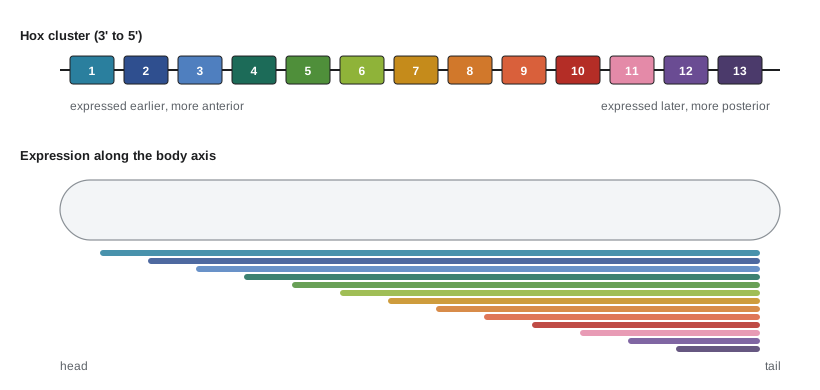
Colinearity seems to depend on keeping the genes together. In flies the clusters have been broken apart in some species without disaster, but in vertebrates the clusters are compact and free of unrelated genes, suggesting strong selection to preserve their organization. Work by Denis Duboule and colleagues has shown that Hox clusters in the mouse are controlled by large regulatory landscapes on either side, megabase-scale regions full of enhancers, that contact the cluster through chromatin looping within topologically associating domains. During trunk development, the cluster appears to open progressively from its 3' end, with genes switching from a repressed chromatin state marked by Polycomb proteins to an active state in sequence. This progressive chromatin opening provides a plausible mechanism for temporal colinearity.
Hox codes and vertebral identity
In vertebrates, Hox genes specify the identity of segments along the axis, most visibly the vertebrae. The mammalian vertebral column is divided into cervical (neck), thoracic (rib-bearing), lumbar, sacral and caudal regions, and almost all mammals, from mice to giraffes, have seven cervical vertebrae. Each region expresses a particular combination of Hox genes, a Hox code. Knockout experiments in mice show the logic. Mice lacking all three Hox10 paralogues (Hoxa10, Hoxc10 and Hoxd10) form ribs on all lumbar vertebrae, which take on a thoracic identity; mice lacking all three Hox11 paralogues have sacral vertebrae transformed to lumbar identity. These are homeotic transformations, the vertebrate equivalent of the fly's four-winged mutants. Because paralogues have partly redundant functions, single mutants often show only mild changes, and the full effect appears only when a whole paralogous group is removed.
When Hox genes are expressed in overlapping domains, the posterior gene generally dominates, a rule called posterior prevalence. A cell expressing Hox genes 1 to 10 adopts the identity specified mainly by Hox10. Posterior prevalence explains why loss of a posterior gene often produces an anterior transformation, as the next most posterior gene takes over, while ectopic expression of a posterior gene in an anterior region produces a posterior transformation.
# A toy model of the vertebrate Hox code with posterior prevalence.
# Each vertebra expresses Hox groups 1..k, where k rises along the axis;
# the identity is set by the most posterior group expressed, mapped to a region.
region_by_group = {4: "cervical", 5: "cervical", 6: "thoracic", 7: "thoracic", 8: "thoracic",
                   9: "thoracic", 10: "lumbar", 11: "sacral", 12: "caudal", 13: "caudal"}
# number of vertebrae with each most-posterior Hox group (illustrative, mouse-like totals)
plan = [(4, 3), (5, 4), (6, 3), (7, 3), (8, 3), (9, 4), (10, 6), (11, 4), (12, 8), (13, 22)]
def column(knockout=None):
    out = []
    for top, n in plan:
        expressed = [g for g in range(1, top + 1) if g != knockout]
        g = max(expressed)                       # posterior prevalence
        out += [region_by_group.get(g, "cervical")] * n
    return out
def summary(col):
    counts = {}
    for r in col: counts[r] = counts.get(r, 0) + 1
    return ", ".join(f"{k} {counts.get(k, 0)}" for k in ("cervical", "thoracic", "lumbar", "sacral", "caudal"))
print("wild type          :", summary(column()))
print("Hox10 group removed:", summary(column(knockout=10)))
print("Hox11 group removed:", summary(column(knockout=11)))wild type : cervical 7, thoracic 13, lumbar 6, sacral 4, caudal 30 Hox10 group removed: cervical 7, thoracic 19, lumbar 0, sacral 4, caudal 30 Hox11 group removed: cervical 7, thoracic 13, lumbar 10, sacral 0, caudal 30
The toy model reproduces the direction of the knockout phenotypes: without Hox10, lumbar vertebrae become thoracic; without Hox11, sacral vertebrae become lumbar. Real Hox codes are more complex. Several paralogous groups act at each level, expression boundaries differ between tissues, and Hox proteins act with cofactors such as Pbx and Meis, which modify their DNA-binding specificity. But the basic logic, that a combination of Hox genes with posterior dominance specifies regional identity, holds broadly.
Hox genes and evolutionary change
If Hox genes specify body regions, changes in their expression should change body plans, and they do. In snakes, the trunk is long and uniform, with rib-bearing vertebrae extending almost to the cloaca; Hox expression domains that mark the thoracic region in limbed vertebrates are expanded along the body. In 2016, Evgeny Kvon, Axel Visel and colleagues showed that snakes have lost function in the ZRS, a limb enhancer of Sonic hedgehog lying within an intron of a distant gene, Lmbr1. Replacing the mouse ZRS with the python version produced mice with severely truncated limbs, and the snake ZRS has a deletion in a region needed for its activity. Changes in the expression boundaries of Hox genes also correlate with the position of the forelimbs in birds and mammals: the boundary between cervical and thoracic vertebrae, and the forelimb, lie at the anterior boundary of Hoxc6 expression in species with very different neck lengths, from frogs with short necks to geese with long ones.
Arthropods provide some of the clearest examples. In insects, the Hox gene Ultrabithorax (Ubx) suppresses leg formation on abdominal segments by repressing the leg gene Distal-less. In crustaceans, which have legs on many segments, Ubx protein lacks the domain needed to repress Distal-less. Changes in the Ubx protein itself thus contributed to the evolution of the insect body plan, with legs restricted to the thorax. In butterflies, Ubx expression in the hindwing makes its pattern different from the forewing.
Deep homology
Some developmental genes control the formation of structures that evolved independently in different lineages. The eyes of insects and vertebrates have long been considered a textbook case of convergence: compound eyes and camera eyes differ in almost every respect. Yet in 1995 Walter Gehring's group showed that expressing the fly gene eyeless in the imaginal discs of legs and wings produced ectopic compound eyes on those appendages, and that the mouse homologue Pax6 could do the same. Mutations in Pax6 cause small eyes in mice (Small eye) and aniridia in humans, and Pax6 is required for eye development in animals from planarians to squid. Neil Shubin, Cliff Tabin and Sean Carroll coined the term deep homology for this situation, in which structures that are not homologous as structures nonetheless use homologous genetic circuits inherited from a common ancestor. A simple light-sensing organ controlled by Pax6 in an ancient ancestor may have been elaborated independently into the eyes of modern animals.
| Example | Developmental gene or element | Change | Evolutionary outcome |
|---|---|---|---|
| Snake limb loss | ZRS enhancer of Sonic hedgehog | Deletion within the enhancer | Loss of limb development |
| Stickleback pelvic reduction | Pitx1 pelvic enhancer | Deletion of a tissue-specific enhancer | Loss of pelvic spines in freshwater populations |
| Insect abdomen without legs | Ultrabithorax protein | Gain of a Distal-less repression domain | Legs restricted to the thorax |
| Butterfly eyespots | Distal-less and other wing patterning genes | Recruitment to new expression domains | Novel wing pattern elements |
| Fruit fly wing spots | yellow gene enhancers | Gain of new binding sites | Male-specific wing pigmentation in some species |
| Beak shape in Darwin's finches | Bmp4 and calmodulin expression | Changes in level and timing | Deeper or longer beaks |
Regulatory evolution
A recurring finding of evo-devo is that much evolutionary change occurs in the regulatory regions of genes rather than in the proteins they encode. The developmental toolkit genes are pleiotropic: each acts in many tissues and stages. A mutation that changes the protein would affect all of them and is likely to be harmful. A mutation in one modular enhancer, by contrast, changes expression in only one place, reducing pleiotropic side effects. The threespine stickleback, discussed in BIOL-321 lesson 10, is the best-known case: freshwater populations in lakes across British Columbia, Alaska and elsewhere repeatedly lost their pelvic spines through deletions of a pelvic enhancer of Pitx1, leaving the gene's other functions intact. Another is the evolution of wing spots in Drosophila species, where Sean Carroll's group showed that new enhancers of the pigmentation gene yellow evolved by gaining binding sites for existing transcription factors.
This does not mean coding changes never matter: Ubx in insects shows they can. Gene duplication also provides raw material, as the vertebrate Hox clusters illustrate; after duplication, paralogues can divide ancestral functions or acquire new ones. And changes in the timing of developmental events, heterochrony, can produce dramatic effects. The axolotl, a salamander that becomes sexually mature while retaining larval features such as gills, is an example of paedomorphosis caused by low thyroid hormone signaling; treating axolotls with thyroid hormone induces metamorphosis.
Novelty and constraint
Evo-devo also asks why some forms evolve repeatedly and others never. Development may constrain evolution, channelling variation along certain paths. The near universality of seven cervical vertebrae in mammals, despite huge variation in neck length, may reflect constraint: changes in cervical number in humans are associated with childhood cancers and stillbirth, suggesting that mutations altering Hox expression in this region have pleiotropic costs. Sloths and manatees, the exceptions, have low metabolic rates, which may relax the constraint. On the other side, new structures can arise by co-opting existing networks: the turtle shell, insect wings, butterfly eyespots and the feathers of birds all appear to have recruited pre-existing developmental pathways to new places.
Worked problem
Interpreting a mouse knockout
A mouse homozygous for a loss-of-function mutation in Hoxc8 has a 14th pair of ribs on the first lumbar vertebra and its first lumbar vertebra resembles the last thoracic vertebra. Suppose a second, hypothetical mouse expresses Hoxc8 throughout the cervical region and develops ribs on the seventh cervical vertebra. Explain both results using posterior prevalence.
Show solution
- In the knockout, a vertebra that normally expresses Hoxc8 as its most posterior active gene now has a less posterior gene dominating, so it takes on a more anterior identity.
- Hoxc8 is normally active in posterior thoracic and upper lumbar regions, so the first lumbar vertebra is transformed toward a thoracic identity and forms ribs.
- In the gain-of-function mouse, Hoxc8 is the most posterior gene expressed in cervical vertebrae, so it dominates and transforms them toward a thoracic identity.
- The seventh cervical vertebra, closest to the thorax, forms ribs.
Answer: Loss of a Hox gene causes an anterior transformation and ectopic expression causes a posterior transformation, both consistent with posterior prevalence. Such results showed that vertebrate Hox genes, like fly homeotic genes, specify regional identity.
Common mistakes
MistakeThinking vertebrates have a single Hox cluster like flies.
InsteadMammals have four clusters, HoxA to HoxD, produced by genome duplications, with 39 genes.
MistakeAssuming colinearity means genes are transcribed one after another as a single unit.
InsteadEach gene is transcribed separately; colinearity refers to the match between gene order, expression position and timing.
MistakeExpecting single Hox gene knockouts to cause dramatic transformations.
InsteadParalogues are partly redundant; complete transformations usually require removing a whole paralogous group.
MistakeCalling eyes in flies and mice homologous structures because both use Pax6.
InsteadThe eyes themselves evolved independently; Pax6 illustrates deep homology of the underlying genetic circuit.
MistakeAssuming most morphological evolution comes from new genes.
InsteadMuch change comes from altered regulation of existing toolkit genes, especially in modular enhancers.
MistakeSaying snakes lost limbs because they lost the Sonic hedgehog gene.
InsteadThey lost function in a limb-specific enhancer (the ZRS); Shh is still used elsewhere.
Summary and key terms
- Hox genes, containing the homeobox, are conserved from flies to humans; mammals have four clusters with 39 genes in 13 paralogous groups.
- Colinearity matches the order of Hox genes on the chromosome to their expression along the body and, in vertebrates, to their timing.
- Large regulatory landscapes and progressive chromatin opening help control Hox clusters.
- Hox codes with posterior prevalence specify vertebral identity; knockouts of paralogous groups cause homeotic transformations.
- Changes in Hox expression and target regulation underlie snake body plans, insect leg loss and forelimb position.
- Pax6 controls eye development across animals, an example of deep homology.
- Much evolutionary change arises from regulatory mutations in modular enhancers, which limit pleiotropy.
- evo-devo
- Evolutionary developmental biology, the study of how changes in development produce evolutionary change.
- Hox genes
- Homeobox-containing transcription factor genes, clustered in the genome, that specify regional identity along the body axis.
- paralogous group
- Hox genes in different clusters derived from the same ancestral gene.
- colinearity
- The correspondence between Hox gene order and expression position and timing along the body axis.
- Hox code
- The combination of Hox genes expressed in a region that specifies its identity.
- posterior prevalence
- The dominance of more posterior Hox genes over anterior ones where they are coexpressed.
- regulatory landscape
- A large chromosomal region containing enhancers that control a gene or gene cluster.
- ZRS
- A long-range limb enhancer of Sonic hedgehog whose loss of function contributed to limb loss in snakes.
- deep homology
- The use of homologous genetic circuits to build structures that evolved independently.
- Pax6
- A transcription factor required for eye development across animals.
- pleiotropy
- The effect of one gene on several different traits.
- heterochrony
- An evolutionary change in the timing of developmental events.
Practice problems
Work each problem on paper first, then reveal the answer to check.
Why do mammals have four Hox clusters?
Show answer
Two rounds of whole-genome duplication early in vertebrate evolution copied the ancestral cluster.
Describe spatial and temporal colinearity.
Show answer
Genes at the 3' end are expressed more anteriorly (spatial) and earlier (temporal) than genes at the 5' end.
Why might single Hox gene knockouts show only mild phenotypes?
Show answer
Paralogues in other clusters have overlapping functions and compensate.
What phenotype do mice lacking all Hox10 paralogues show?
Show answer
Lumbar vertebrae take on a thoracic identity and form ribs.
State the rule of posterior prevalence.
Show answer
Where several Hox genes are coexpressed, the most posterior one generally determines identity.
How does Ubx differ between insects and crustaceans, and why does it matter?
Show answer
Insect Ubx can repress Distal-less, suppressing abdominal legs; crustacean Ubx cannot.
What experiment demonstrated deep homology in eye development?
Show answer
Ectopic expression of fly eyeless or mouse Pax6 in fly imaginal discs produced ectopic compound eyes.
Why are enhancer mutations a common route to evolutionary change?
Show answer
They alter expression in one tissue without the pleiotropic effects of changing the protein everywhere.
How did the stickleback lose its pelvic spines?
Show answer
Deletion of a pelvic enhancer of Pitx1 removed expression in the pelvic region only.
Give an example of heterochrony.
Show answer
The axolotl reaches sexual maturity while retaining larval features because of low thyroid hormone signaling.
Lesson quiz · 5 questions
Score 60% or more to complete this lesson.
Create a free account to save your progress, take quizzes and earn a certificate.