
BIOL-360 · Lesson 6 of 12 · 27 min
Signaling pathways in development
One of the great surprises of modern developmental biology is how few signaling pathways an animal uses to build itself. A handful of families, Wnt, Hedgehog, TGF-β (including BMP and Nodal), receptor tyrosine kinases (especially FGF), Notch and nuclear hormone receptors such as the retinoic acid receptor, account for most of the cell–cell communication in every animal embryo studied, from sea anemones to humans. The same pathway may specify the neural tube in one place, pattern the limb in another and control the growth of hair follicles in a third. BIOL-214 lesson 11 described the receptors and intracellular steps of several of these pathways. This lesson asks a different question: how do developing tissues use them? We look at how signals are produced and spread, how cells interpret their level and duration, how pathways are wired into feedback loops, and what happens when they go wrong.
A small toolkit
Each pathway has a recognizable core. A secreted or membrane-bound ligand binds a receptor; a transduction cascade carries the signal to the nucleus; and a transcription factor changes gene expression. What makes these pathways suitable for development is that their outputs are typically switch-like changes in transcription that can be maintained or reinforced, and that they can be reused by connecting the same core to different target genes in different cells. The specificity lies largely in the responding cell, in its competence, the transcription factors and chromatin state that determine which enhancers respond.
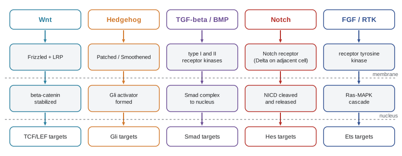
| Pathway | Ligands (examples) | Key transducer | Developmental roles (examples) |
|---|---|---|---|
| Wnt/β-catenin | Wnt1, Wnt3a, Wnt8, Wingless | β-catenin with TCF/LEF | Dorsal axis in frogs, posterior identity, limb outgrowth, segment polarity |
| Hedgehog | Sonic, Indian and Desert hedgehog | Gli transcription factors | Ventral neural tube, limb digit pattern, bone growth plate |
| TGF-β family | BMP2/4/7, Nodal, activin, Dpp | Smad2/3 (Nodal) or Smad1/5/8 (BMP) with Smad4 | Mesoderm induction, dorsal–ventral patterning, left–right asymmetry |
| Receptor tyrosine kinase | FGF4/8/10, EGF | Ras–MAPK, PI3K | Limb outgrowth, posterior mesoderm, somite wavefront |
| Notch | Delta, Serrate/Jagged | Notch intracellular domain with CSL | Lateral inhibition, boundaries, segmentation clock |
| Nuclear receptors | Retinoic acid, thyroid hormone | Ligand-activated transcription factors | Hindbrain patterning, limb, metamorphosis |
Making and moving a signal
For a morphogen to form a gradient, it must be secreted and must move from cell to cell. That is not straightforward for some developmental signals. Hedgehog proteins are modified with cholesterol at their C-terminus and palmitate at their N-terminus, and Wnts are modified with palmitoleic acid by the enzyme Porcupine; these lipids make the proteins hydrophobic and membrane associated. Releasing them requires dedicated machinery, such as Dispatched for Hedgehog and Wntless for Wnt, and they may travel bound to carrier proteins, in lipoprotein particles or on exosomes. Some signals are carried along long, thin cellular extensions called cytonemes, which can contact cells several cell diameters away. Heparan sulfate proteoglycans in the extracellular matrix bind many ligands and shape their distribution, and mutants lacking the enzymes that make them disrupt Hedgehog, Wnt and BMP gradients in flies.
How far a signal spreads also depends on how quickly it is removed. Receptors capture ligands and internalize them, and in some cases this receptor-mediated degradation is the main way the gradient is shaped. Patched, the Hedgehog receptor, is itself a target of Hedgehog signaling: cells that receive Hedgehog make more Patched, which binds and sequesters Hedgehog, restricting its further spread. This negative feedback steepens the gradient and makes it more robust to changes in Hedgehog production. Secreted antagonists, such as the BMP inhibitors of lesson 5 and the Wnt inhibitors Dickkopf and the secreted Frizzled-related proteins, add another layer of control.
Reading the level: Sonic hedgehog in the neural tube
The vertebrate neural tube provides a classic example of a morphogen read by concentration and time. Sonic hedgehog (Shh) is secreted first by the notochord beneath the neural tube and then by the floor plate, the ventral midline of the tube itself. It forms a ventral-to-dorsal gradient. Progenitor cells exposed to the highest levels become floor plate; slightly lower levels specify progenitors of V3 interneurons, then motor neurons, then V2, V1 and V0 interneurons at progressively more dorsal positions. Dorsal fates are promoted by BMP and Wnt from the roof plate. Cells read the gradient through Gli transcription factors: without Hedgehog, Gli proteins are processed into repressors; with Hedgehog, full-length activators predominate. Each progenitor domain is defined by a combination of transcription factors such as Nkx2.2, Olig2, Nkx6.1, Pax6 and Irx3.
Two features of this system are important. First, cells integrate the signal over time: in experiments by James Briscoe and colleagues, neural progenitors exposed to a given concentration of Shh moved to more ventral identities the longer they were exposed, and the intracellular response adapts, as Patched upregulation reduces sensitivity. Second, the transcription factors induced by Shh cross-repress one another: Nkx2.2 and Pax6 repress each other, as do Olig2 and Irx3. This cross-repression converts a smooth gradient into sharp boundaries between progenitor domains and makes the decisions stable once made, much as the gap genes do in Drosophila.
Notch and lateral inhibition
The Notch pathway is unusual because both ligand and receptor are membrane proteins, so signaling occurs only between cells in contact. When Delta on one cell binds Notch on an adjacent cell, Notch is cleaved twice, first by an ADAM metalloprotease and then by γ-secretase, releasing the Notch intracellular domain, which enters the nucleus and, with the DNA-binding protein CSL, activates target genes such as the Hes repressors. One widespread use of Notch is lateral inhibition. In a field of initially equivalent cells, any cell that by chance expresses slightly more Delta activates Notch more strongly in its neighbors, which then reduce their own Delta expression and adopt a secondary fate. The result is a salt-and-pepper pattern of cells adopting the primary fate surrounded by cells adopting the secondary fate. In the Drosophila neuroectoderm, lateral inhibition singles out neuroblasts; mutations in Notch or Delta cause too many cells to become neural, the neurogenic phenotype after which the pathway's genes were named. In vertebrates, lateral inhibition spaces sensory hair cells in the inner ear and endocrine cells in the gut and pancreas.
import random
random.seed(3)
# Lateral inhibition on a hexagonal-like grid (Collier et al. 1996 model, simplified).
# dn/dt = f(mean adjacent-cell Delta) - n ;  dd/dt = g(n) - d
# f rises with adjacent cell Delta (Notch activation); g falls with Notch (Delta repressed).
R, C = 8, 12
n = [[random.uniform(0.0, 0.1) for _ in range(C)] for _ in range(R)]
d = [[random.uniform(0.9, 1.0) for _ in range(C)] for _ in range(R)]
def nbrs(i, j):
    off = [(-1, 0), (1, 0), (0, -1), (0, 1)] + ([(-1, -1), (1, -1)] if i % 2 == 0 else [(-1, 1), (1, 1)])
    return [((i + a) % R, (j + b) % C) for a, b in off]
f = lambda x: x ** 2 / (0.01 + x ** 2)          # Notch activation by adjacent cells' Delta
g = lambda x: 1 / (1 + 100 * x ** 2)            # Delta repression by Notch
dt = 0.1
for step in range(3000):
    mean_d = [[sum(d[a][b] for a, b in nbrs(i, j)) / 6 for j in range(C)] for i in range(R)]
    n = [[n[i][j] + dt * (f(mean_d[i][j]) - n[i][j]) for j in range(C)] for i in range(R)]
    d = [[d[i][j] + dt * (g(n[i][j]) - d[i][j]) for j in range(C)] for i in range(R)]
primary = 0
for i in range(R):
    row = ''.join('#' if d[i][j] > 0.5 else '.' for j in range(C))
    primary += row.count('#')
    print((' ' if i % 2 else '') + ' '.join(row))
print(f"high-Delta (primary fate) cells: {primary} of {R * C} = {100 * primary / (R * C):.0f}%")# . . # . . # . . # . . . # . . # . . # . . # . # . . # . . # . . # . . . # . . # . . # . . # . # . . # . . # . . # . . . # . . # . . # . . # . # . . # . . # . . # . . . # . . # . . # . . # . high-Delta (primary fate) cells: 32 of 96 = 33%
Starting from nearly uniform cells with small random differences, the simulation settles into a pattern in which isolated high-Delta cells (marked #), the cells adopting the primary fate, are surrounded by low-Delta cells. No cell is told its fate in advance; the pattern emerges from feedback between adjacent cells. This is a second kind of self-organization alongside the reaction–diffusion systems of lesson 1, and the mathematical model, published by Joanne Collier and colleagues in 1996, showed that the feedback must be sufficiently steep to produce a pattern. Notch is also used for lateral induction, in which signaling spreads a fate rather than suppressing it, in boundary formation, as at the dorsal–ventral boundary of the fly wing, and in the segmentation clock of lesson 9.
FGF, retinoic acid and opposing gradients
Fibroblast growth factors act through receptor tyrosine kinases and the Ras–MAPK cascade. In development they promote proliferation and outgrowth, as in the limb bud and the tail bud, and they help maintain cells in an undifferentiated state. FGF8 in the posterior of the vertebrate embryo forms a gradient that keeps posterior cells in a progenitor state. Retinoic acid, a small lipid-soluble derivative of vitamin A made by the enzyme Raldh2 in somites and degraded by Cyp26 enzymes, forms an opposing gradient from more anterior tissues and promotes differentiation. The two antagonize each other, so that the boundary between them moves posteriorly as the embryo elongates, marking where somites and neurons will form. Retinoic acid acts through nuclear retinoic acid receptors bound to target genes, including Hox genes, and so specifies the identity of hindbrain segments; this is why excess vitamin A is a teratogen, as lesson 12 explains.
Feedback, crosstalk and robustness
Developmental pathways rarely act alone. They are wired into networks with recurring motifs. Negative feedback, as in the Hedgehog pathway's induction of Patched, the Wnt pathway's induction of its inhibitor Axin2 and the BMP pathway's induction of inhibitory Smads, limits signaling and stabilizes gradients. Positive feedback, as in the Wingless–Hedgehog loop of lesson 4 or Nodal's induction of its own expression, locks in decisions and creates bistability. Pathways also intersect: Wnt and BMP cooperate to specify the neural crest, FGF and BMP antagonize each other in the neural plate, and Shh in the limb maintains FGF expression in the apical ectoderm. Understanding development increasingly means modeling these networks quantitatively.
When pathways fail
Because the same pathways are used repeatedly, mutations in them cause characteristic syndromes affecting several organs. Mutations in human SHH cause holoprosencephaly, failure of the forebrain to divide into two hemispheres, which in its most severe form produces a single central eye (cyclopia). The same defect occurs in lambs whose mothers eat corn lily (Veratrum californicum), which contains cyclopamine, an alkaloid that blocks the Hedgehog transducer Smoothened; this connection was traced in the 1950s and 1960s after outbreaks of cyclopic lambs in Idaho. Mutations in FGF receptors cause skeletal dysplasias: a single recurrent mutation in FGFR3, which makes the receptor overactive, causes achondroplasia, the most common form of short-limbed dwarfism, and other FGFR mutations cause craniosynostosis, the premature fusion of skull sutures. Notch pathway mutations cause Alagille syndrome, affecting the liver, heart and skeleton.
The same pathways that build embryos also maintain adult tissues and are frequently reactivated in cancer. Mutations in the Wnt pathway inhibitor APC initiate most colorectal cancers by stabilizing β-catenin in intestinal stem cells. Loss of the Hedgehog receptor Patched causes basal cell carcinoma and medulloblastoma, and Smoothened inhibitors derived from the study of cyclopamine, vismodegib and sonidegib, are approved for advanced basal cell carcinoma. The connection between developmental biology and cancer runs throughout modern biomedicine, and is the subject of BIOL-450.
Worked problem
Reading a neural tube experiment
Chick neural plate explants are cultured with different concentrations of Shh protein for 24 hours. At 0.5 nM they express Pax7 (a dorsal marker); at 2 nM they express Pax6 and some Olig2; at 4 nM they express Olig2 and Nkx6.1 (motor neuron progenitors); at 8 nM they express Nkx2.2. A second set of explants at 2 nM is cultured for 48 hours instead of 24, and now expresses Olig2 and Nkx6.1. What do the results show about how cells read Shh?
Show solution
- Different concentrations produce different ventral identities, so cells respond to Shh concentration, consistent with a morphogen.
- Higher concentrations produce more ventral identities, matching the normal ventral source.
- At the same concentration, longer exposure produces a more ventral identity, so cells also integrate the duration of signaling.
- Cross-repression between transcription factors such as Pax6 and Nkx2.2 would sharpen these responses into discrete domains.
Answer: Neural progenitors read both the concentration and the duration of Shh signaling. A cell's identity reflects its cumulative exposure, which is one reason why gradients that change over time can still give precise patterns.
Common mistakes
MistakeAssuming each developmental event uses its own dedicated pathway.
InsteadA small set of pathways is reused many times; specificity comes from the responding cell.
MistakeThinking morphogens simply diffuse freely.
InsteadLipid-modified Wnts and Hedgehogs need dedicated release, carriers, proteoglycans or cytonemes, and receptors shape their spread.
MistakeTreating Notch as a long-range morphogen.
InsteadNotch signaling needs direct cell contact because Delta and Notch are both membrane proteins.
MistakeBelieving a cell reads only the concentration of a morphogen.
InsteadDuration of exposure and adaptation also matter, as in the neural tube.
MistakeAssuming negative feedback weakens a pattern.
InsteadNegative feedback, such as Patched induction, often makes gradients steeper and more robust.
MistakeSeparating developmental genes from cancer genes.
InsteadMany oncogenes and tumor suppressors, such as APC and Patched, are components of developmental pathways.
Summary and key terms
- A few pathway families (Wnt, Hedgehog, TGF-β, RTK/FGF, Notch, nuclear receptors) are reused throughout development.
- Specificity comes from the competence of the responding cell, its transcription factors and chromatin.
- Lipid-modified signals need dedicated release and transport; proteoglycans, receptors and antagonists shape gradients.
- Shh patterns the ventral neural tube through Gli activators and repressors, read by concentration and duration and sharpened by cross-repression.
- Delta–Notch lateral inhibition produces spaced primary-fate cells from an equivalent field.
- Opposing FGF and retinoic acid gradients define where posterior tissues differentiate.
- Pathway mutations cause syndromes such as holoprosencephaly and achondroplasia and are reactivated in cancer.
- Sonic hedgehog
- A secreted Hedgehog family protein that patterns the ventral neural tube and the limb.
- Gli
- Transcription factors that mediate Hedgehog signaling, acting as activators or repressors.
- Patched
- The Hedgehog receptor, which inhibits Smoothened in the absence of ligand.
- floor plate
- The ventral midline of the neural tube, a source of Sonic hedgehog.
- cytoneme
- A thin cellular extension that carries signaling proteins between cells.
- lateral inhibition
- A process by which a cell adopting a fate prevents its neighbors from adopting the same fate.
- Delta
- A membrane-bound ligand that activates Notch on adjacent cells.
- Notch intracellular domain
- The fragment released by cleavage of activated Notch that enters the nucleus to regulate genes.
- retinoic acid
- A vitamin A derivative that acts through nuclear receptors to pattern the hindbrain and other tissues.
- holoprosencephaly
- Failure of the forebrain to divide into two hemispheres, often caused by defects in Hedgehog signaling.
- cyclopamine
- A plant alkaloid that blocks Smoothened and causes cyclopia in embryos.
- achondroplasia
- A form of dwarfism caused by an activating mutation in FGFR3.
Practice problems
Work each problem on paper first, then reveal the answer to check.
Why can the same pathway produce different outcomes in different tissues?
Show answer
The responding cells differ in competence: transcription factors and chromatin determine which genes respond.
How does Patched induction shape the Hedgehog gradient?
Show answer
Cells receiving Hedgehog make more Patched, which binds and sequesters Hedgehog, limiting its spread and steepening the gradient.
Which cells in the neural tube receive the highest Shh, and what do they become?
Show answer
The most ventral cells next to the floor plate; they become floor plate and V3 progenitors.
What is the significance of cross-repression between Nkx2.2 and Pax6?
Show answer
It converts a graded Shh input into a sharp, stable boundary between progenitor domains.
What phenotype results from loss of Notch in the fly neuroectoderm, and why?
Show answer
Too many neuroblasts (neurogenic phenotype), because lateral inhibition fails.
In the simulation, what produces the initial differences between cells?
Show answer
Small random variation in starting Delta and Notch levels, amplified by feedback.
How do FGF and retinoic acid interact in the posterior embryo?
Show answer
They form opposing gradients that antagonize each other, defining where differentiation begins.
Why did lambs born in Idaho in the 1950s have a single eye?
Show answer
Their mothers ate corn lily containing cyclopamine, which blocks Hedgehog signaling and prevents forebrain division.
Explain why achondroplasia is dominant.
Show answer
A gain-of-function FGFR3 mutation makes the receptor overactive, so one copy is enough to inhibit bone growth.
Give an example of a developmental pathway gene mutated in cancer.
Show answer
APC in colorectal cancer, or Patched in basal cell carcinoma.
Lesson quiz · 5 questions
Score 60% or more to complete this lesson.
Create a free account to save your progress, take quizzes and earn a certificate.