
BIOC-200 · Lesson 8 of 12 · 24 min
Nucleic acids
▶ Video coming soon A video version of this lesson is on the way.
Nucleic acids store and transmit the information that makes each organism what it is. Deoxyribonucleic acid, DNA, holds the genetic instructions in a remarkably stable form and copies them faithfully every time a cell divides. Ribonucleic acid, RNA, carries copies of those instructions to the ribosomes, helps translate them into proteins, and in many cases catalyzes reactions or regulates genes itself. This lesson describes the chemical units of nucleic acids, the structure of the double helix, the differences between DNA and RNA, and the chemistry that makes nucleic acids so suited to their job. It closes with the flow of information from DNA to RNA to protein, which BIOC-340 covers in detail.
Nucleotides
Nucleic acids are polymers of nucleotides. Each nucleotide has three parts: a five-carbon sugar, a nitrogen-containing base and one or more phosphate groups. In RNA the sugar is ribose; in DNA it is 2-deoxyribose, which lacks the hydroxyl group on carbon 2. To avoid confusion with the atoms of the base, the carbons of the sugar are numbered with primes, 1′ to 5′. The base is attached to the 1′ carbon by a glycosidic bond, and the phosphate to the 5′ carbon. A sugar with a base but no phosphate is called a nucleoside; adding phosphates gives a nucleotide, such as adenosine monophosphate (AMP), adenosine diphosphate (ADP) or adenosine triphosphate (ATP).
The bases come in two families. The purines, adenine (A) and guanine (G), have two fused rings. The pyrimidines, cytosine (C), thymine (T) and uracil (U), have a single ring. DNA contains A, G, C and T; RNA contains A, G, C and U. Uracil differs from thymine only by lacking a methyl group. The bases are flat, partly aromatic and only slightly soluble in water, and they absorb ultraviolet light strongly near 260 nm, which is how the concentration of DNA and RNA is usually measured.
Nucleotides do much more than build nucleic acids. ATP is the energy currency of the cell, as Lesson 9 explains, and GTP powers protein synthesis and signaling. Cyclic AMP, made from ATP, carries hormone signals inside cells. Several coenzymes are built around nucleotides: NAD⁺, FAD and coenzyme A all contain an adenosine unit, a hint, some biochemists think, of an early stage of life in which RNA did most of the work.
The phosphodiester backbone
Nucleotides are joined by phosphodiester bonds. The phosphate on the 5′ carbon of one nucleotide links to the hydroxyl on the 3′ carbon of the next, so the backbone is a chain of alternating sugar and phosphate, with the bases sticking out to the side. Like a polypeptide, a nucleic acid strand has direction. One end carries a free 5′ phosphate and the other a free 3′ hydroxyl, and sequences are written from the 5′ end to the 3′ end. The sequence 5′-ATGC-3′ is therefore different from 5′-CGTA-3′. Each phosphate in the backbone carries a negative charge at physiological pH, so nucleic acids are large polyanions; in cells their charges are balanced by magnesium ions, polyamines and positively charged proteins such as histones.
The double helix
By 1950 Erwin Chargaff had found a striking regularity in DNA from many species: the amount of adenine always equalled the amount of thymine, and the amount of guanine equalled the amount of cytosine, although the ratio of A + T to G + C varied from species to species. X-ray diffraction photographs of DNA fibers taken by Rosalind Franklin and Raymond Gosling at King's College London, particularly the famous Photograph 51, showed that DNA is a helix with a regular repeat. In 1953 James Watson and Francis Crick, at Cambridge, combined these clues with model building and proposed the double helix. Watson, Crick and Maurice Wilkins received the 1962 Nobel Prize in Physiology or Medicine; Franklin had died in 1958, and the prize is not awarded after death.
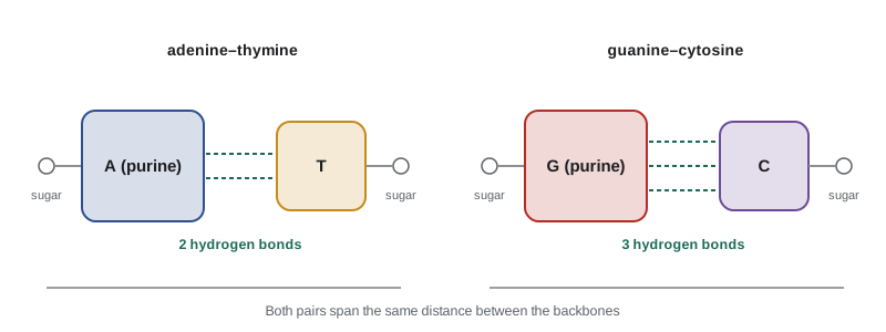
In the Watson–Crick model two strands wind around a common axis in a right-handed helix, running in opposite directions: they are antiparallel. The sugar-phosphate backbones lie on the outside, facing the water, and the bases are stacked flat inside, perpendicular to the axis, like the steps of a spiral staircase. Each base on one strand pairs with a base on the other by hydrogen bonds: adenine with thymine through two hydrogen bonds, and guanine with cytosine through three. Each pair joins a purine to a pyrimidine, so every base pair is the same width and fits neatly between the backbones, and the pairing explains Chargaff's rules exactly.
In the common B form of DNA, the helix is about 2.0 nm wide, the base pairs are 0.34 nm apart, and there are about 10.5 base pairs per turn, giving a repeat of about 3.6 nm. Because the sugars of a base pair are not directly opposite each other, the backbones form two grooves of different sizes along the helix, the major groove and the minor groove. Proteins that recognize particular DNA sequences, such as the transcription factors that switch genes on, usually read the edges of the bases exposed in the major groove without unwinding the helix.
The two strands are complementary: the sequence of one determines the sequence of the other. If one strand reads 5′-ATGCTT-3′, its partner must read 5′-AAGCAT-3′, written in the conventional 5′-to-3′ direction. Watson and Crick noted at once that this specific pairing suggests a copying mechanism. When the strands separate, each can serve as a template for a new partner, and in 1958 Matthew Meselson and Franklin Stahl showed that DNA is copied in exactly this semiconservative way, each new double helix containing one old strand and one new.
What holds the helix together
Hydrogen bonds between base pairs give the double helix its specificity, but they are not the main source of its stability. In water, the bases could just as well form hydrogen bonds with water molecules. Much of the stability comes from base stacking: the flat bases above and below each other attract through van der Waals forces and the hydrophobic effect, which buries their faces away from water. Stacking between G–C pairs is especially favorable, and together with their third hydrogen bond this makes G–C-rich DNA more stable than A–T-rich DNA. Repulsion between the negatively charged phosphates works against the helix, which is why DNA is more stable in salt solutions, where positive ions screen the charges.
Heating DNA separates its strands, a process called melting or denaturation. Because stacked bases absorb less ultraviolet light than unstacked ones, the absorbance at 260 nm rises by about 40% as the strands come apart, which makes melting easy to follow. The temperature at which half the DNA is melted is its melting temperature, Tm. It rises with the G–C content and with the salt concentration. On cooling, complementary strands find each other again and re-form the helix, a process called annealing or hybridization. Hybridization is the basis of many laboratory methods: the polymerase chain reaction, PCR, uses short single strands called primers that anneal to the ends of a target sequence, and DNA microarrays and many diagnostic tests detect sequences by hybridization to complementary probes.
RNA
RNA differs from DNA in three ways that change its behavior. Its sugar is ribose, whose 2′ hydroxyl group can attack the neighboring phosphodiester bond under basic conditions, so RNA is much less chemically stable than DNA. It uses uracil instead of thymine. And it is usually single-stranded. A single strand of RNA folds back on itself wherever complementary stretches can pair, forming short double-helical stems with loops at their ends, and these hairpins fold further into complex three-dimensional shapes, much as a polypeptide folds into a protein. RNA also tolerates base pairs outside the Watson–Crick rules, the most common being the G–U wobble pair.
Cells make several kinds of RNA. Messenger RNA (mRNA) carries the sequence of a gene from the DNA to the ribosomes, where it is read three bases at a time to specify the amino acids of a protein. Transfer RNA (tRNA), about 76 nucleotides folded into an L shape, carries each amino acid to the ribosome and matches it to the right three-base codon. Ribosomal RNA (rRNA) forms the structural and catalytic core of the ribosome; the peptidyl transferase center that joins amino acids is made of RNA, so the ribosome is a ribozyme. Many other RNAs, including microRNAs of about 22 nucleotides, regulate which genes are used. Because RNA can both carry information and catalyze reactions, many biochemists think that early life passed through an RNA world before DNA and proteins took over their specialized roles.
Why DNA uses deoxyribose and thymine
The small chemical differences between DNA and RNA suit each to its role. Removing the 2′ hydroxyl makes DNA hundreds of times more resistant to hydrolysis, which is why it can store information for a lifetime, and why fragments of DNA can survive for tens of thousands of years in bones and permafrost. The use of thymine solves a problem of repair. Cytosine slowly loses its amino group in a reaction called deamination, which turns it into uracil. If DNA normally contained uracil, a repair enzyme could not tell which uracils were mistakes. Because DNA uses thymine, any uracil in DNA is recognized as damage and removed by the enzyme uracil-DNA glycosylase. RNA, which is short-lived and copied from DNA as needed, does not need this protection.
The flow of genetic information
Crick summarized the relationship between nucleic acids and proteins in 1958 as the central dogma of molecular biology: information flows from DNA to RNA by transcription and from RNA to protein by translation, and once information has passed into protein it cannot get back out. DNA is copied by DNA polymerases during replication. RNA polymerases copy genes into RNA. Ribosomes translate mRNA into protein using the genetic code, in which each of 64 three-base codons specifies one of the twenty amino acids or a stop signal. Retroviruses such as HIV add a reverse step, copying their RNA genomes into DNA with the enzyme reverse transcriptase, which is why the dogma is now stated as a flow of information rather than a strict one-way street.
The human genome contains about 3.1 billion base pairs in each set of chromosomes and roughly 20,000 protein-coding genes, which together make up less than 2% of the DNA. If the DNA of the 46 chromosomes in one cell were stretched out, it would be about 2 meters long, yet it fits into a nucleus about 6 µm across, wrapped around histone proteins into structures called nucleosomes and folded further into chromatin.
A worked example: base composition and melting
Worked problem
Using Chargaff's rules and a melting-temperature estimate
A sample of double-stranded DNA contains 22% adenine. (a) What are the proportions of T, G and C? (b) A short double-stranded DNA of 20 base pairs has the sequence 5′-ATGCGTACCGGATTACGCAT-3′ on one strand. Use the simple rule Tm = 2 °C × (number of A–T pairs) + 4 °C × (number of G–C pairs), often used for short primers, to estimate its melting temperature.
Show solution
- Chargaff: A = T, so T = 22%. Together A + T = 44%, so G + C = 56%, and G = C = 28%.
- Count the bases of the 20-mer: A appears 5 times, T 5 times, G 5 times and C 5 times. So there are 10 A–T pairs and 10 G–C pairs.
- Tm ≈ 2 × 10 + 4 × 10 = 60 °C.
- The rule reflects the extra stability of G–C pairs, which contribute twice as much in this approximation; real melting temperatures also depend on salt, DNA concentration and the exact order of the bases.
Answer: (a) T = 22%, G = 28% and C = 28%. (b) About 60 °C.
A program: working with sequences
Sequence analysis is one of the most common tasks in biochemistry, and much of it can be done with a few lines of code. The program finds the complementary strand of a DNA sequence in the conventional 5′-to-3′ direction, calculates its G–C content and a simple melting temperature, transcribes it into RNA, and translates the RNA into protein with the genetic code, stopping at the first stop codon.
bases = "TCAG"
codons = [a + b + c for a in bases for b in bases for c in bases]
amino = "FFLLSSSSYY**CC*WLLLLPPPPHHQQRRRRIIIMTTTTNNKKSSRRVVVVAAAADDEEGGGG"
CODE = dict(zip(codons, amino))     # the standard genetic code; * = stop

def reverse_complement(dna):
    pair = {"A": "T", "T": "A", "G": "C", "C": "G"}
    return "".join(pair[b] for b in reversed(dna))

def gc_content(dna):
    return (dna.count("G") + dna.count("C")) / len(dna)

def wallace_tm(dna):
    at = dna.count("A") + dna.count("T")
    return 2 * at + 4 * (len(dna) - at)

def translate(dna):
    protein = []
    for i in range(0, len(dna) - 2, 3):
        aa = CODE[dna[i:i + 3]]
        if aa == "*":
            break
        protein.append(aa)
    return "".join(protein)

gene = "ATGGCTAGCAAAGGAGAAGAACTTTTCACTGGAGTTGTCCCAATTCTTGTTGAATTAGATTAA"
print("coding strand  5'-" + gene + "-3'")
print("template       5'-" + reverse_complement(gene) + "-3'")
print(f"length {len(gene)} bp, GC content {gc_content(gene):.1%}")
print("mRNA           5'-" + gene.replace("T", "U") + "-3'")
print("protein        " + translate(gene))
primer = "ATGCGTACCGGATTACGCAT"
print()
print(f"primer {primer}: GC {gc_content(primer):.0%}, Tm about {wallace_tm(primer)} C")coding strand 5'-ATGGCTAGCAAAGGAGAAGAACTTTTCACTGGAGTTGTCCCAATTCTTGTTGAATTAGATTAA-3' template 5'-TTAATCTAATTCAACAAGAATTGGGACAACTCCAGTGAAAAGTTCTTCTCCTTTGCTAGCCAT-3' length 63 bp, GC content 36.5% mRNA 5'-AUGGCUAGCAAAGGAGAAGAACUUUUCACUGGAGUUGUCCCAAUUCUUGUUGAAUUAGAUUAA-3' protein MASKGEELFTGVVPILVELD primer ATGCGTACCGGATTACGCAT: GC 50%, Tm about 60 C
The gene in the program is the beginning of the coding sequence of green fluorescent protein, the jellyfish protein that glows green and is used everywhere as a marker in cell biology, followed by a stop codon. The translation, MASKGEELFTGVVPILVELD, matches the first residues of the protein, with an extra alanine at position 2 as in many laboratory versions of the gene. The template strand is the one the RNA polymerase reads; the mRNA has the same sequence as the coding strand, with U in place of T. Real genes are much longer, and in eukaryotes they are interrupted by stretches called introns that are spliced out of the RNA before translation, but the logic of complementarity and the genetic code is the same.
Common mistakes
MistakeThe two strands of DNA run in the same direction.
InsteadThey are antiparallel: one runs 5′ to 3′ and its partner 3′ to 5′.
MistakeHydrogen bonds are the main force holding the double helix together.
InsteadThey provide specificity; base stacking contributes most of the stability.
MistakeA–T and G–C pairs are held by the same number of hydrogen bonds.
InsteadA–T pairs have two hydrogen bonds and G–C pairs three.
MistakeRNA is just DNA with uracil.
InsteadRNA also has ribose, whose 2′ hydroxyl makes it less stable, and it is usually single-stranded and folds into complex shapes.
MistakeChargaff's rules mean every DNA has 25% of each base.
InsteadThey mean A = T and G = C within double-stranded DNA; the G + C content varies between species.
MistakeA sequence reads the same written in either direction.
InsteadNucleic acid sequences have direction; 5′-ATGC-3′ differs from 5′-CGTA-3′.
Summary and key terms
- Nucleotides contain a pentose sugar, a purine or pyrimidine base and phosphate; they are linked by 5′–3′ phosphodiester bonds into strands with direction.
- DNA is an antiparallel, right-handed double helix in which A pairs with T and G with C, explaining Chargaff's rules.
- Base stacking provides most of the stability of the helix and hydrogen bonding its specificity; DNA melts on heating and re-anneals on cooling.
- RNA uses ribose and uracil, is usually single-stranded, folds into complex structures and includes mRNA, tRNA, rRNA and regulatory RNAs.
- Information flows from DNA to RNA by transcription and from RNA to protein by translation using the genetic code.
- Nucleotide
- A sugar, a base and one or more phosphate groups; the unit of nucleic acids.
- Purine
- A two-ring base: adenine or guanine.
- Pyrimidine
- A one-ring base: cytosine, thymine or uracil.
- Phosphodiester bond
- The link between the 3′ carbon of one nucleotide and the 5′ carbon of the next through a phosphate.
- Antiparallel
- Running in opposite directions, as the two strands of the double helix do.
- Melting temperature (Tm)
- The temperature at which half of a double-stranded nucleic acid has separated into single strands.
- Codon
- A sequence of three bases in mRNA that specifies an amino acid or a stop signal.
Practice problems
Work each problem on paper first, then reveal the answer to check.
Write the complementary strand of 5′-GATTACA-3′, in the 5′-to-3′ direction.
Show answer
5′-TGTAATC-3′.
A double-stranded DNA is 35% G. What share of the bases is A?
Show answer
C = 35%, so G + C = 70% and A = T = 15%.
Which would melt at a higher temperature, a DNA that is 30% G + C or one that is 60% G + C? Why?
Show answer
The 60% G + C DNA, because G–C pairs stack more favorably and have three hydrogen bonds.
How long is a DNA of 10,500 base pairs in the B form, and how many helical turns does it make?
Show answer
10,500 × 0.34 nm = 3.57 µm, and 10,500/10.5 = 1,000 turns.
Why does DNA contain thymine instead of uracil?
Show answer
Deaminated cytosine becomes uracil; using thymine lets repair enzymes recognize any uracil in DNA as damage.
What is the difference between a nucleoside and a nucleotide?
Show answer
A nucleotide has one or more phosphate groups; a nucleoside is only the sugar and base.
Lesson quiz · 5 questions
Score 60% or more to complete this lesson.
Create a free account to save your progress, take quizzes and earn a certificate.