
BIOL-214 · Lesson 8 of 12 · 30 min
The cytoskeleton I: actin and cell motility
A cell is not a bag of fluid with organelles floating in it. Its shape, its ability to move, the positions of its organelles and the separation of its chromosomes all depend on the cytoskeleton, a network of protein filaments that are constantly assembled, disassembled and reorganized. There are three main kinds: actin filaments, microtubules and intermediate filaments. Each is built from many copies of a small subunit, which lets the cell build long structures quickly from a pool of soluble pieces and take them apart just as quickly. BIOL-107 gave an overview of the three systems and of motor proteins. This lesson examines actin filaments in depth: how they assemble and treadmill, how dozens of binding proteins control where and when they grow, how myosin motors pull on them in muscle and other cells, and how a cell crawls across a surface. Lesson 9 covers microtubules, intermediate filaments and microtubule motors.
Actin: subunits and filaments
Actin is one of the most abundant proteins in eukaryotic cells, often 5 to 10 percent of total protein and over 20 percent in muscle. The 42-kDa globular subunit, G-actin, binds a molecule of ATP or ADP in a cleft between its two lobes. Subunits assemble head to tail into a filament, F-actin, about 7 nm in diameter, which can be described as two strands twisting gently around each other. Because all subunits face the same way, the filament has a structural polarity. The two ends are called the barbed (or plus) end and the pointed (or minus) end, names that come from the arrowhead pattern seen in electron micrographs when filaments are decorated with myosin heads. Actin is highly conserved: yeast and human actins are about 89 percent identical, and most eukaryotes cannot survive without it.
Nucleation, elongation and critical concentration
Purified G-actin with ATP and salt polymerizes into filaments, but not immediately. The first step, nucleation, is slow because dimers and trimers of actin are unstable; once a nucleus of three or four subunits forms, elongation is fast. In a test tube, polymerization therefore shows a lag phase, a growth phase and a steady state. At steady state, the concentration of free subunits settles at a value called the critical concentration, C_c. Above C_c filaments grow; below it they shrink.
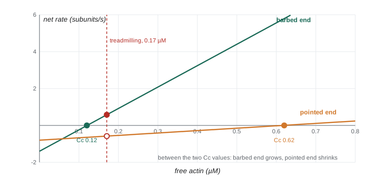
For each end, the rate of addition is proportional to the free subunit concentration, while the rate of loss is constant, so the net rate is k_on × C − k_off and the critical concentration for that end is k_off / k_on. The two ends differ. The barbed end adds and loses subunits much faster, and for ATP-actin its critical concentration is lower than at the pointed end.
| End | k_on (µM⁻¹ s⁻¹) | k_off (s⁻¹) | Critical concentration (µM) |
|---|---|---|---|
| Barbed (plus) end, ATP-actin | 11.6 | 1.4 | 0.12 |
| Pointed (minus) end, ATP-actin | 1.3 | 0.8 | 0.6 |
Inside a filament, each subunit hydrolyzes its bound ATP to ADP a few seconds after joining and releases the phosphate more slowly. Older parts of the filament therefore contain ADP-actin, which binds its neighbors less tightly. Because the barbed end grows fast, it usually has a cap of ATP-actin; the pointed end, where subunits linger longer, is mostly ADP-actin.
Treadmilling
Since the two ends have different critical concentrations, there is a range of free actin concentrations, between 0.12 and 0.6 µM in the table, in which subunits add at the barbed end while they are lost from the pointed end. At one particular concentration, the addition and loss exactly balance, and the filament keeps a constant length while individual subunits move through it from the barbed end to the pointed end, like the tread on a tank. This behavior, called treadmilling, depends on ATP hydrolysis, which provides the energy for the difference between the ends.
# Treadmilling with the rate constants for ATP-actin (subunits per second)
kon_b, koff_b = 11.6, 1.4      # barbed end
kon_p, koff_p = 1.3, 0.8       # pointed end

Cc_b, Cc_p = koff_b / kon_b, koff_p / kon_p
print(f"critical concentration: barbed {Cc_b:.2f} uM, pointed {Cc_p:.2f} uM")

# Treadmilling concentration: barbed gain equals pointed loss
C_tm = (koff_b + koff_p) / (kon_b + kon_p)
flux = kon_b * C_tm - koff_b
print(f"treadmilling at C = {C_tm:.2f} uM; flux = {flux:.2f} subunits/s through each filament")
print(f"that is {flux * 2.7:.1f} nm/s, or {flux * 2.7 * 60 / 1000:.2f} um per minute")

# Simulate filament length at different free actin levels
for C in (0.05, C_tm, 0.4, 1.0):
    net_b, net_p = kon_b*C - koff_b, kon_p*C - koff_p
    print(f"C = {C:4.2f} uM: barbed {net_b:+6.2f}/s, pointed {net_p:+5.2f}/s, "
f"length change {(net_b + net_p)*2.7:+6.1f} nm/s")critical concentration: barbed 0.12 uM, pointed 0.62 uM treadmilling at C = 0.17 uM; flux = 0.58 subunits/s through each filament that is 1.6 nm/s, or 0.09 um per minute C = 0.05 uM: barbed -0.82/s, pointed -0.74/s, length change -4.2 nm/s C = 0.17 uM: barbed +0.58/s, pointed -0.58/s, length change +0.0 nm/s C = 0.40 uM: barbed +3.24/s, pointed -0.28/s, length change +8.0 nm/s C = 1.00 uM: barbed +10.20/s, pointed +0.50/s, length change +28.9 nm/s
Each subunit adds about 2.7 nm to the filament, because the two strands share the length. The calculation shows that pure actin treadmills slowly, about 0.6 subunits per second. In cells, actin turns over hundreds of times faster, and filaments at the leading edge of a crawling cell grow at about 0.1 to 1 µm per second. This speed comes from accessory proteins that change the rates at each end.
Actin-binding proteins control everything
Cells contain a high concentration of unpolymerized actin, often 50 to 200 µM, far above the critical concentration, yet it does not all polymerize. Binding proteins hold it in check and direct polymerization to the right places.
| Protein | Activity | Effect in cells |
|---|---|---|
| Thymosin β4 | Sequesters G-actin | Keeps a large pool of subunits from polymerizing |
| Profilin | Binds G-actin, promotes ADP-to-ATP exchange, delivers subunits to barbed ends | Supports fast growth only at free barbed ends |
| Arp2/3 complex | Nucleates a new filament from the side of an existing one at 70° | Builds branched networks in lamellipodia and endocytic sites |
| Formins | Nucleate and stay on the growing barbed end, speeding elongation | Build long unbranched bundles: filopodia, stress fibers, contractile ring |
| Capping protein | Binds barbed ends and stops growth | Keeps branched networks short and dense |
| Cofilin (ADF) | Binds ADP-actin, severs and depolymerizes filaments | Recycles subunits rapidly from older filaments |
| Fimbrin, α-actinin, filamin | Cross-link filaments | Form tight bundles (microvilli) or loose networks (cortex) |
The Arp2/3 complex, discovered by Thomas Pollard and colleagues, contains two actin-related proteins that mimic an actin dimer and so overcome the barrier to nucleation. It is inactive on its own and is switched on by nucleation-promoting factors such as WASP and the WAVE complex, which are in turn activated by the small GTPases Cdc42 and Rac. Mutations in WASP cause Wiskott–Aldrich syndrome, an immune deficiency in which immune cells cannot rearrange actin properly. Some bacteria hijack the system: Listeria monocytogenes, a cause of food poisoning and serious infections in pregnancy, carries a surface protein that activates Arp2/3, so it propels itself through the host cytoplasm on a comet tail of actin and pushes into neighboring cells without leaving the protection of the cell.
Drugs and toxins that act on actin are powerful research tools. Cytochalasins cap barbed ends; latrunculins sequester G-actin; jasplakinolide stabilizes filaments; and phalloidin, from the death cap mushroom Amanita phalloides, binds and stabilizes F-actin. Because phalloidin binds only filaments, fluorescent phalloidin is the standard stain for F-actin in fixed cells.
Myosins: motors that walk on actin
Myosins are motor proteins that convert the energy of ATP hydrolysis into movement along actin filaments, almost always toward the barbed end. Humans have about 40 myosin genes in more than ten classes. Myosin II, the muscle myosin, has two heavy chains, each with a globular motor head at one end, and a long coiled-coil tail; many myosin II molecules assemble by their tails into bipolar thick filaments with heads at both ends. Myosin V, by contrast, is a two-headed motor that walks along actin hand over hand in steps of 36 nm, carrying vesicles and organelles. Myosin I has a single head and a tail that binds membranes.
The mechanochemical cycle of myosin couples ATP hydrolysis to a change in shape. In the rigor state, a head without nucleotide binds tightly to actin. ATP binding releases the head. Hydrolysis of ATP cocks the lever arm, a long helix stabilized by light chains. The head rebinds actin weakly, then release of phosphate triggers the power stroke, in which the lever arm swings and moves the filament about 5 to 10 nm. Release of ADP returns the head to the rigor state. Without ATP, heads remain locked to actin, which is why muscles stiffen after death in rigor mortis.
Muscle contraction
In skeletal muscle, actin and myosin are arranged in repeating units called sarcomeres, about 2.5 µm long at rest, which give the muscle its striated appearance. Thin filaments of actin are anchored at the Z discs at each end of the sarcomere; thick filaments of myosin II sit in the middle, overlapping the thin filaments. When myosin heads pull the thin filaments toward the center, each sarcomere shortens, while the filaments themselves do not change length: the sliding-filament model, proposed in 1954 by Andrew Huxley with Rolf Niedergerke and by Hugh Huxley with Jean Hanson. A giant elastic protein, titin, spans half a sarcomere and acts as a spring.
Contraction is switched on by calcium. At rest, tropomyosin lies along the thin filament and blocks the myosin-binding sites. When a nerve impulse reaches the muscle fiber, it spreads along the plasma membrane and down into the T tubules, triggering calcium release from the sarcoplasmic reticulum (lesson 3). Calcium binds troponin C, part of a complex attached to tropomyosin, which shifts tropomyosin and exposes the binding sites. When stimulation stops, SERCA pumps calcium back into the sarcoplasmic reticulum and the muscle relaxes. Smooth muscle and non-muscle cells regulate myosin II differently: calcium activates a kinase, myosin light-chain kinase, which phosphorylates the regulatory light chain, and the Rho-activated kinase ROCK increases contraction further.
Worked problem
How fast can a muscle shorten?
A muscle fiber 4 cm long contains sarcomeres 2.5 µm long arranged in series. If each half sarcomere shortens at 1.25 µm per second, how fast does the whole fiber shorten?
Show solution
- Number of sarcomeres in series = 0.04 m / 2.5 × 10⁻⁶ m = 16,000.
- Each sarcomere has two halves, each shortening at 1.25 µm/s, so each sarcomere shortens at 2.5 µm/s.
- Fiber shortening speed = 16,000 × 2.5 µm/s = 40,000 µm/s = 4 cm/s.
- Arranging small units in series multiplies speed; arranging fibers in parallel multiplies force.
Answer: About 4 cm/s, the fiber’s own length each second (illustrative values).
Mutations in sarcomere proteins cause cardiomyopathies and muscular dystrophies. Duchenne muscular dystrophy, which affects about 1 in 3,500 to 5,000 boys, results from loss of dystrophin, a large protein that links the actin cytoskeleton of muscle fibers through the membrane to the extracellular matrix (lesson 10); without it, fibers are damaged during contraction and gradually replaced by fat and scar tissue. Hypertrophic cardiomyopathy, often caused by mutations in cardiac myosin, is a common cause of sudden death in young athletes. A myosin inhibitor, mavacamten, approved in the United States in 2022 and later in other countries, reduces excessive contraction in this disease.
How cells crawl
Many cells move by crawling across surfaces: fibroblasts healing a wound, white blood cells hunting bacteria, neurons extending their growth cones and, harmfully, cancer cells invading tissue. Crawling can be broken into steps that are coordinated in time and space.
| Step | Structures and molecules | Controlled mainly by |
|---|---|---|
| Protrusion of the leading edge | Lamellipodia (branched Arp2/3 networks), filopodia (formin-built bundles) | Rac (lamellipodia), Cdc42 (filopodia) |
| Adhesion at the front | Integrins cluster into focal adhesions linked to actin by talin and vinculin | Integrin activation; force |
| Contraction and translocation of the cell body | Myosin II pulling on actin stress fibers and the cortex | Rho and its kinase ROCK |
| De-adhesion at the rear | Release or breakage of adhesions; proteolysis | Calcium, calpain, myosin tension |
At the leading edge, actin polymerization itself generates the pushing force. Filaments in the lamellipodium are oriented with their barbed ends toward the membrane. As thermal motion briefly opens a gap between the membrane and a filament tip, a subunit adds, and the filament prevents the membrane from moving back, a mechanism called a Brownian ratchet. Meanwhile, the whole network moves backward relative to the substrate, in retrograde flow, as myosin pulls on it and subunits are disassembled further back by cofilin. Where the network is gripped by adhesions to the substrate, retrograde flow slows and protrusion is converted into forward motion, like a car’s clutch engaging. Keratocytes, fish skin cells, can move at 10 to 15 µm per minute with almost unchanging shape, which made them a favorite model for this mechanism.
The Rho family GTPases, Rho, Rac and Cdc42, act as master regulators. Experiments in which active forms of each were injected into fibroblasts, carried out by Alan Hall and colleagues in the 1990s, showed that Rho produces stress fibers and focal adhesions, Rac produces lamellipodia and Cdc42 produces filopodia. In a moving cell, these activities are segregated: Rac and Cdc42 are active at the front, Rho at the rear. Chemical gradients sensed by receptors can steer the cell by shifting where these GTPases are active, a process called chemotaxis.
import numpy as np
rng = np.random.default_rng(214)

# Persistent random walk: tracks of crawling cells imaged every minute (simulated)
def track(speed, persistence, steps=120):
    angle, x, y, path = rng.uniform(0, 2*np.pi), 0.0, 0.0, []
    for _ in range(steps):
        angle += rng.normal(0, persistence)       # turning noise, radians per minute
        x += speed*np.cos(angle); y += speed*np.sin(angle); path.append((x, y))
    return np.array(path)

for label, speed, turn in [("keratocyte-like", 12.0, 0.1), ("fibroblast-like", 1.0, 0.6),
                           ("highly random", 1.0, 2.0)]:
    p = track(speed, turn)
    total = speed * len(p)
    net = np.hypot(*p[-1])
    print(f"{label:16s} speed {speed:4.1f} um/min: path {total:6.0f} um, "
f"net displacement {net:6.0f} um, straightness {net/total:.2f}")keratocyte-like speed 12.0 um/min: path 1440 um, net displacement 1073 um, straightness 0.74 fibroblast-like speed 1.0 um/min: path 120 um, net displacement 12 um, straightness 0.10 highly random speed 1.0 um/min: path 120 um, net displacement 5 um, straightness 0.04
Migration is usually summarized by speed and by straightness, the net displacement divided by the total path length. Cells with high persistence, like keratocytes, travel nearly straight; fibroblasts wander. Treating a cell with a drug that blocks Rac or Arp2/3, or depleting a protein by RNA interference, and then measuring these quantities from time-lapse images is a standard experiment in cell biology and cancer research.
Actin in other jobs
Actin does far more than drive crawling and contraction. A thin layer of cross-linked actin and myosin under the plasma membrane, the cortex, gives animal cells their shape and stiffness. At the end of mitosis, a contractile ring of actin and myosin II, assembled by formins under the control of Rho, pinches the cell in two (lesson 12). Bundles of actin cross-linked by fimbrin and villin support the microvilli of intestinal cells, increasing their absorptive surface, and the stereocilia of inner-ear hair cells, whose deflection by sound opens mechanosensitive channels. Actin assembly helps pinch off endocytic vesicles, drives phagocytosis and moves some organelles. Even the nucleus contains actin, which takes part in transcription and DNA repair.
Common mistakes
MistakeActin filaments are permanent structures.
InsteadThey turn over continuously; in cells, accessory proteins accelerate assembly and disassembly.
MistakeBoth ends of an actin filament grow at the same rate.
InsteadThe barbed end grows much faster and has a lower critical concentration than the pointed end.
MistakeMyosin II walks along actin carrying vesicles.
InsteadMyosin II forms filaments that contract actin networks; myosin V and others carry cargo.
MistakeIn muscle contraction, actin and myosin filaments shorten.
InsteadThey slide past each other; the sarcomere shortens while filament lengths stay constant.
MistakeArp2/3 builds long parallel bundles.
InsteadArp2/3 builds branched networks; formins build long unbranched bundles.
Summary and key terms
- Actin subunits bind ATP and assemble into polar filaments with fast-growing barbed and slow pointed ends.
- Nucleation is slow; the critical concentration k_off/k_on sets whether ends grow or shrink.
- Different critical concentrations at the two ends, powered by ATP hydrolysis, allow treadmilling.
- Profilin, thymosin, Arp2/3, formins, capping protein and cofilin control where and how fast actin assembles.
- Myosins convert ATP hydrolysis into movement; myosin II drives contraction and myosin V carries cargo.
- Muscle contracts by filament sliding in sarcomeres, switched on by calcium binding to troponin.
- Cells crawl by protrusion, adhesion, contraction and de-adhesion, coordinated by Rac, Cdc42 and Rho.
- G-actin
- The globular actin subunit, which binds ATP or ADP.
- barbed end
- The fast-growing plus end of an actin filament.
- critical concentration
- The free subunit concentration at which net growth at an end is zero.
- treadmilling
- Steady addition at one end and loss at the other, so subunits flow through a filament of constant length.
- Arp2/3 complex
- A complex that nucleates branched actin filaments from the sides of existing ones.
- formin
- A protein that nucleates unbranched actin filaments and remains on the growing barbed end.
- cofilin
- A protein that severs and depolymerizes ADP-actin filaments.
- sarcomere
- The repeating contractile unit of striated muscle.
- lamellipodium
- A broad, flat protrusion at the front of a crawling cell, filled with branched actin.
- Rho GTPases
- The small GTPases Rho, Rac and Cdc42 that organize the actin cytoskeleton.
Practice problems
Work each problem on paper first, then reveal the answer to check.
Why does purified actin polymerization show a lag phase?
Show answer
Nucleation is slow because small actin oligomers are unstable.
Calculate the critical concentration for an end with k_on = 10 µM⁻¹s⁻¹ and k_off = 2 s⁻¹.
Show answer
2/10 = 0.2 µM.
At a free actin concentration of 0.3 µM, does the pointed end grow or shrink (C_c = 0.6 µM)?
Show answer
It shrinks, because 0.3 µM is below its critical concentration.
What is the role of profilin?
Show answer
It binds G-actin, promotes exchange of ADP for ATP and delivers subunits to free barbed ends.
How does Listeria move inside host cells?
Show answer
Its surface protein activates Arp2/3, building an actin comet tail that pushes the bacterium.
Why is fluorescent phalloidin used to stain actin?
Show answer
It binds specifically to F-actin, not G-actin.
What causes rigor mortis?
Show answer
Without ATP, myosin heads stay bound to actin, locking the filaments.
How does calcium switch on skeletal muscle contraction?
Show answer
It binds troponin C, which shifts tropomyosin away from myosin-binding sites on actin.
Which Rho GTPase produces lamellipodia, and which produces stress fibers?
Show answer
Rac produces lamellipodia; Rho produces stress fibers.
A cell moves along a path of 200 µm but ends 50 µm from its start. What is its straightness?
Show answer
50/200 = 0.25.
Lesson quiz · 5 questions
Score 60% or more to complete this lesson.
Create a free account to save your progress, take quizzes and earn a certificate.