
BIOL-214 · Lesson 10 of 12 · 30 min
Cell junctions, adhesion and the extracellular matrix
Cells in an animal body are not isolated units. They stick to one another and to the material around them, seal off compartments, exchange small molecules directly and sense mechanical forces through their attachments. The proteins that make these connections link the cytoskeleton of one cell to that of its neighbors or to the extracellular matrix, the network of secreted proteins and polysaccharides that fills the spaces between cells. Together they turn individual cells into tissues that can bear loads, form barriers and keep their shape. This lesson describes the junctions that join cells, the adhesion molecules behind them, the major components of the extracellular matrix and how cells use adhesion to sense their surroundings. It also examines how the breakdown of adhesion contributes to cancer spread, a theme developed in BIOL-450.
A map of cell junctions
Epithelia, the sheets of cells that line body surfaces and cavities, show all the main junction types in a characteristic order from the apical surface toward the base of the cell. Each type has a distinct job.
| Junction | Main transmembrane proteins | Linked to | Function |
|---|---|---|---|
| Tight junction | Claudins, occludin | Actin (via ZO proteins) | Seals the space between cells; separates apical and basolateral membranes |
| Adherens junction | E-cadherin (classical cadherins) | Actin (via catenins) | Cell–cell adhesion; transmits tension along a sheet |
| Desmosome | Desmogleins, desmocollins (cadherins) | Intermediate filaments (keratins) | Strong spot welds that resist mechanical stress |
| Gap junction | Connexins (vertebrates) | None | Channels that pass ions and small molecules between cells |
| Hemidesmosome | Integrin α6β4, collagen XVII | Intermediate filaments | Anchors cells to the basal lamina |
| Focal adhesion | Integrins | Actin (via talin, vinculin) | Anchors cells to the matrix; sensing and signaling |
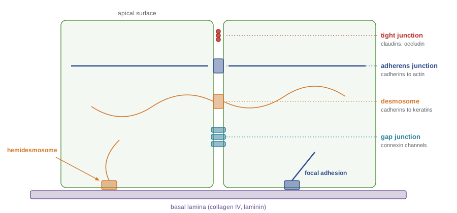
Tight junctions and barriers
Tight junctions form a belt around the top of each epithelial cell where strands of claudin and occludin in neighboring membranes meet and nearly close the space between them. They do two things. First, they act as a barrier, controlling what passes between cells (the paracellular pathway). Second, they act as a fence, preventing membrane proteins and lipids in the outer leaflet from diffusing between the apical and basolateral domains, which keeps the polarized distribution of transporters needed for absorption and secretion (lesson 7).
Tight junctions are not equally tight everywhere. Humans have about 27 claudin genes, and the particular mixture of claudins expressed by an epithelium decides how leaky it is and which ions can pass. Claudin-2, for example, forms pores for cations and water in the leaky proximal tubule of the kidney, while claudin-16 and claudin-19 allow magnesium and calcium to pass in the thick ascending limb; mutations in them cause a rare inherited magnesium-wasting disease. Some bacterial toxins exploit claudins: the enterotoxin of Clostridium perfringens, a common cause of food poisoning, binds claudin-3 and claudin-4 and damages the intestinal lining.
Barrier tightness can be measured as transepithelial electrical resistance (TEER). Cells are grown as a sheet on a porous filter insert, electrodes are placed on each side, and the resistance to a small current is measured. Leaky epithelia have TEER values of tens of Ω·cm², while tight ones, such as the bladder lining or the blood–brain barrier formed by brain endothelial cells, reach thousands.
import numpy as np
rng = np.random.default_rng(214)

# TEER: subtract the blank filter, then multiply by the membrane area
area_cm2 = 1.12                  # a 12-well insert
blank = 120.0                    # ohms, filter without cells
days = np.arange(1, 9)
true_teer = 1200 / (1 + np.exp(-(days - 4.5)))          # ohm.cm2 rising as junctions form
measured = blank + true_teer / area_cm2 + rng.normal(0, 15, days.size)
teer = (measured - blank) * area_cm2
for d, m, t in zip(days, measured, teer):
    print(f"day {d}: reading {m:6.0f} ohm -> TEER {t:6.0f} ohm.cm2")

# A treatment that opens tight junctions (simulated day 8 values, triplicate)
control = np.array([1150, 1210, 1180.0])
treated = np.array([340, 410, 375.0])
print(f"treatment lowers TEER by {100*(1 - treated.mean()/control.mean()):.0f}%")day 1: reading 178 ohm -> TEER 65 ohm.cm2 day 2: reading 206 ohm -> TEER 96 ohm.cm2 day 3: reading 331 ohm -> TEER 237 ohm.cm2 day 4: reading 520 ohm -> TEER 448 ohm.cm2 day 5: reading 778 ohm -> TEER 737 ohm.cm2 day 6: reading 1006 ohm -> TEER 993 ohm.cm2 day 7: reading 1117 ohm -> TEER 1116 ohm.cm2 day 8: reading 1158 ohm -> TEER 1162 ohm.cm2 treatment lowers TEER by 68%
Two details matter in practice. The resistance of an empty filter must be subtracted, and the result must be multiplied by the membrane area, not divided, because a larger sheet offers more parallel paths and so a lower resistance for the same tissue. Researchers use TEER to follow the formation of barrier layers, to test drugs that must cross the intestine or the blood–brain barrier, and to study how inflammatory signals loosen junctions in conditions such as inflammatory bowel disease.
Cadherins and adherens junctions
Cadherins are the main cell–cell adhesion molecules of animal tissues. A classical cadherin, such as E-cadherin in epithelia, N-cadherin in neurons and muscle or VE-cadherin in blood vessel endothelium, has five repeated extracellular domains held rigid by calcium ions bound between them. Without calcium the extracellular region becomes floppy and adhesion fails, which is why calcium-free solutions containing EDTA are used to detach cells from culture dishes. Cadherins on opposing cells bind mostly to the same type of cadherin (homophilic binding). In classic experiments by Malcolm Steinberg and Masatoshi Takeichi, cells expressing different cadherins, or different amounts of the same cadherin, sorted themselves out from mixtures, much as oil and water separate, showing how adhesion can help build tissues during development.
Inside the cell, the cadherin tail binds β-catenin and p120-catenin, and β-catenin binds α-catenin, which links to actin filaments. In epithelia, adherens junctions form a continuous belt, and contraction of the actin–myosin belt can fold a whole sheet, for instance as the neural tube forms. β-Catenin also has a second life as a transcriptional regulator in the Wnt signaling pathway (lesson 11), which means that the amount of β-catenin held at junctions can influence gene expression.
Desmosomes and hemidesmosomes
Desmosomes are button-like junctions in which the desmosomal cadherins desmoglein and desmocollin connect, through plakoglobin, plakophilin and desmoplakin, to keratin intermediate filaments. The keratin networks of neighboring cells are thereby linked into a single tissue-wide web, which is why tissues that face mechanical stress, such as skin and heart muscle, are rich in desmosomes. Desmosomal diseases make the point vividly. In pemphigus vulgaris, the immune system produces antibodies against desmoglein 3 (and often desmoglein 1); skin and mucous membrane cells separate and form fragile blisters. Mutations in desmosomal proteins cause arrhythmogenic cardiomyopathy, in which heart muscle cells pull apart under stress and are replaced by fat and fibrous tissue.
Hemidesmosomes, by contrast, anchor the base of epithelial cells to the basal lamina, linking keratin filaments through integrin α6β4 and collagen XVII to laminin in the matrix. Their failure causes junctional and other forms of epidermolysis bullosa, in which the top layer of skin separates from the layer beneath (lesson 9).
Gap junctions
Gap junctions connect the cytoplasm of neighboring cells directly. Each cell contributes a hemichannel, the connexon, built from six connexin subunits, and two connexons dock across the narrow gap to form a channel with a pore about 1.5 nm wide. Ions, second messengers such as cyclic AMP and IP₃, and metabolites up to roughly 1 kDa can pass, but proteins and nucleic acids cannot. Gap junctions cluster into plaques of hundreds or thousands of channels.
Gap junctions couple cells electrically and metabolically. In the heart, they let the action potential spread rapidly from cell to cell so that the muscle contracts in a coordinated wave; in the lens of the eye, which has no blood supply, they distribute nutrients; and between some neurons, they form fast electrical synapses. Channels close when calcium rises or the pH falls, isolating damaged cells from their neighbors. Humans have 21 connexin genes, and mutations in connexin 26 are the most common cause of inherited non-syndromic hearing loss, because the channels are needed to recycle potassium in the cochlea. Plants have an analogous but structurally different connection, the plasmodesma, a membrane-lined channel through the cell wall.
The extracellular matrix
The extracellular matrix (ECM) is made by the cells it surrounds. Its composition varies enormously, from the hard, mineralized matrix of bone to the transparent matrix of the cornea and the thin basal lamina beneath every epithelium. Most matrices are built from the same families of molecules.
| Component | Structure | Main role |
|---|---|---|
| Fibrillar collagens (types I, II, III) | Triple helices of (Gly-X-Y) repeats assembled into fibrils | Tensile strength in skin, tendon, bone and cartilage |
| Collagen IV | Flexible triple helices that form a sheet-like network | Scaffold of the basal lamina |
| Elastin | Cross-linked, highly disordered protein | Elastic recoil in arteries, lungs and skin |
| Glycosaminoglycans and proteoglycans | Long, negatively charged sugar chains, mostly attached to core proteins | Attract water and resist compression; bind growth factors |
| Fibronectin | Dimer with binding sites for integrins, collagen and heparin | Links cells to the matrix; guides migration in development and healing |
| Laminins | Cross-shaped trimers | Key adhesive component of the basal lamina |
Collagen is the most abundant protein in mammals, about a quarter of all protein. Each collagen molecule is a triple helix of three polypeptide chains wound around one another. Every third residue in the helical region must be glycine, because only glycine, with a single hydrogen atom as its side chain, fits in the crowded center of the helix. Many of the other positions are proline and hydroxyproline. Hydroxylation of proline, which takes place in the ER (lesson 3), requires vitamin C; without it, collagen helices are unstable and the result is scurvy, with bleeding gums, poor wound healing and fragile blood vessels. Collagen is secreted as procollagen with extension peptides that keep it soluble; these are trimmed outside the cell, and the molecules then assemble into fibrils that are cross-linked by lysyl oxidase.
# Check collagen-like sequences for the Gly-X-Y repeat
def gly_xy_check(seq, start=0):
    bad = [i for i in range(start, len(seq), 3) if seq[i] != "G"]
    pro_x = sum(seq[i+1] == "P" for i in range(start, len(seq)-1, 3))
    hyp_y = sum(seq[i+2] in "PO" for i in range(start, len(seq)-2, 3))
    return bad, pro_x, hyp_y

# A stretch based on the alpha1(I) chain (O = hydroxyproline), and a mutant form
normal = "GPOGPAGPOGEOGKPGERGAOGPOGPAGPOGERGPOGPO"
mutant = normal[:15] + "S" + normal[16:]       # a glycine replaced by serine

for name, s in [("normal", normal), ("mutant", mutant)]:
    bad, px, hy = gly_xy_check(s)
    print(f"{name}: {len(s)//3} triplets; non-glycine at residue index {bad}; "
f"Pro at X {px}, Pro/Hyp at Y {hy}")normal: 13 triplets; non-glycine at residue index []; Pro at X 8, Pro/Hyp at Y 9 mutant: 13 triplets; non-glycine at residue index [15]; Pro at X 8, Pro/Hyp at Y 9
Replacing a single glycine with a bulkier residue, as in the mutant, kinks the helix. Because each collagen molecule contains three chains, one mutant chain can spoil molecules that also contain normal chains, which is called a dominant negative effect. Such glycine substitutions in type I collagen cause the most severe forms of osteogenesis imperfecta, or brittle bone disease. Mutations in other collagens and in the enzymes that process them cause forms of Ehlers–Danlos syndrome, with stretchy skin and loose joints, while mutations in fibrillin, a protein of elastic fibers, cause Marfan syndrome.
Glycosaminoglycans (GAGs) are long unbranched chains of repeating disaccharides, often sulfated, that carry many negative charges. Hyaluronan is a huge GAG made at the cell surface without a protein core; the others, such as chondroitin sulfate and heparan sulfate, are attached to core proteins to form proteoglycans. Their charges attract cations and water, forming a hydrated gel that resists compression. In cartilage, the proteoglycan aggrecan forms enormous aggregates on hyaluronan, which is why cartilage can bear the weight of the body. Heparan sulfate proteoglycans at the cell surface and in the basal lamina also bind growth factors such as FGF, controlling where signals act.
Integrins: linking matrix to cytoskeleton
Integrins are the main receptors that bind cells to the matrix. Each is a heterodimer of an α and a β subunit; humans have 18 α and 8 β subunits, which combine into 24 integrins with different ligand preferences. Many integrins recognize the short sequence Arg-Gly-Asp (RGD) found in fibronectin and other matrix proteins. Inside the cell, the β subunit binds talin and kindlin, which link to actin. Integrins can switch between a bent, low-affinity shape and an extended, high-affinity shape. Signals inside the cell, such as those that activate white blood cells, cause talin and kindlin to bind and switch integrins on (inside-out signaling); conversely, ligand binding and clustering trigger signals inside the cell (outside-in signaling), including activation of focal adhesion kinase (FAK) and Src.
The ability to switch integrins on and off is vital. White blood cells roll along blood vessel walls until a chemokine signal activates their integrin LFA-1, which then binds firmly to ICAM-1 on the vessel wall so the cells can stop and squeeze out into infected tissue. In leukocyte adhesion deficiency, missing β2 integrins or kindlin-3 leave patients unable to fight bacterial infections. Platelets activate their integrin αIIbβ3 to bind fibrinogen and form clots, and drugs that block this integrin prevent clotting during some heart procedures. BIOL-380 returns to leukocyte adhesion in the context of immune responses.
Mechanotransduction
Cells feel the mechanical properties of their surroundings through their adhesions. When a focal adhesion is pulled by myosin, talin unfolds and exposes new binding sites for vinculin, strengthening the link. Cells on stiff surfaces build large focal adhesions and stress fibers; cells on soft surfaces do not. These differences change gene expression: the transcriptional co-activators YAP and TAZ move into the nucleus when cells are on stiff matrix or spread out and stay in the cytoplasm when cells are on soft matrix or crowded. In a widely cited experiment, Dennis Discher and colleagues showed in 2006 that mesenchymal stem cells grown on soft gels like brain tissue tended toward neuron-like fates, those on intermediate stiffness toward muscle and those on stiff gels toward bone. Matrix stiffening in tumors and fibrotic organs feeds back on cell behavior and is an active area of cancer research.
Worked problem
Interpreting an adhesion-blocking experiment
Fibroblasts are plated on fibronectin-coated wells in medium containing a peptide with the sequence GRGDS, or a control peptide GRGES (in which aspartate is replaced by glutamate). After 30 minutes, 85 percent of control-peptide cells are attached and spread, but only 12 percent of GRGDS-treated cells. What do you conclude?
Show solution
- The GRGDS peptide contains the RGD motif recognized by many integrins.
- The soluble peptide competes with fibronectin for the integrin binding site.
- The control peptide, differing by a single methylene group, has little effect, so the inhibition is specific.
- Most of the adhesion to fibronectin therefore depends on RGD-binding integrins such as α5β1 and αvβ3.
Answer: Adhesion to fibronectin is mediated mainly by RGD-recognizing integrins (illustrative data).
Adhesion and cancer
Most cancers arise in epithelia, and as they become invasive, their cells often lose E-cadherin and gain N-cadherin and vimentin, a shift called epithelial–mesenchymal transition (EMT). Cells that have undergone EMT are less bound to their neighbors and more able to migrate. Germline mutations in the E-cadherin gene, CDH1, cause hereditary diffuse gastric cancer, and many carriers choose to have their stomach removed preventively. Invading tumor cells also secrete matrix metalloproteinases that degrade basement membranes. Integrin signaling helps tumor cells survive outside their normal location, because most epithelial cells undergo a form of programmed death called anoikis when they lose contact with the matrix (lesson 12). BIOL-450 examines invasion and metastasis in detail.
Common mistakes
MistakeTight junctions are equally tight in every epithelium.
InsteadTheir claudin composition makes some epithelia leaky and others very tight.
MistakeTEER is calculated by dividing the resistance by the filter area.
InsteadSubtract the blank filter and multiply by the area to get Ω·cm².
MistakeCadherins work without calcium.
InsteadCalcium rigidifies the extracellular domains; removing it abolishes adhesion.
MistakeDesmosomes link to actin filaments.
InsteadDesmosomes link to intermediate filaments; adherens junctions link to actin.
MistakeGlycine in collagen can be replaced by any small amino acid without harm.
InsteadOnly glycine fits at the center of the triple helix; substitutions cause disease such as osteogenesis imperfecta.
Summary and key terms
- Epithelia contain tight junctions, adherens junctions, desmosomes, gap junctions, hemidesmosomes and focal adhesions.
- Tight junctions made of claudins and occludin seal the paracellular route and act as a fence; TEER measures their tightness.
- Calcium-dependent cadherins provide homophilic cell–cell adhesion and connect to actin through catenins.
- Desmosomes and hemidesmosomes connect intermediate filaments for strength; their loss causes blistering diseases.
- Gap junctions built from connexins let ions and small molecules pass directly between cells.
- The ECM contains collagens, elastin, proteoglycans, fibronectin and laminins; collagen needs glycine every third residue.
- Integrins bind matrix ligands such as RGD, switch between activity states and transmit mechanical signals.
- tight junction
- A junction of claudins and occludin that seals the space between epithelial cells.
- TEER
- Transepithelial electrical resistance, a measure of barrier tightness in Ω·cm².
- cadherin
- A calcium-dependent cell–cell adhesion protein that usually binds the same cadherin type.
- desmosome
- A junction linking the intermediate filament networks of adjacent cells.
- gap junction
- A cluster of connexin channels connecting the cytoplasm of neighboring cells.
- basal lamina
- A thin sheet of matrix, rich in collagen IV and laminin, beneath epithelia.
- collagen
- A family of matrix proteins built as triple helices of Gly-X-Y repeats.
- proteoglycan
- A core protein carrying glycosaminoglycan chains.
- integrin
- An α/β heterodimeric receptor linking the matrix to the cytoskeleton.
- mechanotransduction
- The conversion of mechanical forces into biochemical signals.
Practice problems
Work each problem on paper first, then reveal the answer to check.
What two roles do tight junctions play?
Show answer
A barrier to paracellular passage and a fence separating apical and basolateral membrane domains.
A filter with cells reads 820 Ω, the blank reads 120 Ω and the area is 0.33 cm². What is the TEER?
Show answer
(820 − 120) × 0.33 = 231 Ω·cm².
Why does EDTA help detach cultured cells?
Show answer
It removes calcium, so cadherins lose their rigid shape and adhesion fails.
Which protein links cadherins to actin?
Show answer
α-Catenin, bound to β-catenin.
What is attacked in pemphigus vulgaris?
Show answer
Desmoglein 3 (and often desmoglein 1) in desmosomes, by autoantibodies.
How large a molecule can pass through a gap junction?
Show answer
Up to roughly 1 kDa.
Why does vitamin C deficiency weaken connective tissue?
Show answer
Prolyl hydroxylase needs vitamin C; without hydroxyproline, collagen helices are unstable.
In a 30-residue collagen stretch beginning with glycine, at which positions must glycine occur?
Show answer
Positions 1, 4, 7, … 28, every third residue (10 glycines).
What does inside-out signaling do to integrins?
Show answer
Talin and kindlin bind the β tail and switch the integrin to its high-affinity extended form.
What changes in cadherin expression accompany EMT?
Show answer
Loss of E-cadherin and gain of N-cadherin.
Lesson quiz · 5 questions
Score 60% or more to complete this lesson.
Create a free account to save your progress, take quizzes and earn a certificate.