
BIOL-214 · Lesson 3 of 12 · 32 min
The endoplasmic reticulum and protein synthesis on membranes
The endoplasmic reticulum (ER) is the largest membrane system in most eukaryotic cells: a continuous network of flattened sacs and branching tubules that extends from the nuclear envelope to the cell periphery and encloses a single internal space, the lumen. In a liver cell it accounts for about half of all membrane. The ER is the entry point to the secretory pathway. Nearly every protein that ends up in the plasma membrane, the Golgi apparatus, endosomes or lysosomes, or that is secreted from the cell, begins its journey by crossing or inserting into the ER membrane. The ER is also where most membrane lipids are made, where proteins are folded, glycosylated and checked before they leave, and where calcium is stored for signaling. BIOL-207 introduced the signal recognition particle and the idea of sorting signals; this lesson follows proteins into and through the ER in detail, and examines what happens when the ER’s folding capacity is overwhelmed.
Rough and smooth ER
Under the electron microscope, parts of the ER are studded with ribosomes, giving the rough ER its name, while other parts are bare. Rough ER sheets dominate in cells that secrete large amounts of protein, such as pancreatic acinar cells, plasma cells making antibodies, and liver cells making serum albumin. Smooth ER tubules are abundant in cells specialized for lipid metabolism: steroid-hormone-producing cells of the adrenal cortex and gonads, liver cells that detoxify drugs, and muscle cells, where a specialized smooth ER called the sarcoplasmic reticulum stores the calcium that triggers contraction. The two regions are continuous; ribosomes are not permanent parts of the membrane but attach only while they are making proteins with ER-targeting signals.
| Function | Mainly in | Key molecules or processes |
|---|---|---|
| Co-translational import and insertion of proteins | Rough ER | SRP, SRP receptor, Sec61 translocon, signal peptidase |
| N-linked glycosylation | Rough ER | Oligosaccharyltransferase adds a 14-sugar precursor to Asn |
| Folding, disulfide bonds, quality control | ER lumen | BiP, calnexin, calreticulin, protein disulfide isomerase |
| Lipid synthesis | Smooth ER (and rough ER) | Phospholipids, cholesterol, ceramide |
| Detoxification | Smooth ER of liver | Cytochrome P450 enzymes |
| Calcium storage and release | ER and sarcoplasmic reticulum | SERCA pumps, IP₃ and ryanodine receptors |
| Contacts with other organelles | ER contact sites | Lipid and calcium transfer to mitochondria and plasma membrane |
Co-translational translocation
A protein destined for the secretory pathway starts life on a free ribosome in the cytosol. When its N-terminal signal peptide emerges from the ribosome exit tunnel, the signal recognition particle binds it and briefly slows translation. The SRP brings the whole ribosome–nascent chain complex to the SRP receptor on the ER membrane. Both SRP and its receptor are GTPases; when they meet and hydrolyze their GTP, the ribosome is handed to the Sec61 translocon, a protein-conducting channel, and SRP is released to work again. Translation resumes, and the growing chain is threaded through the channel into the lumen as it is made. Signal peptidase on the luminal side cuts off the signal peptide. Because the ribosome sits tightly on the channel, the polypeptide never contacts the cytosol, and its synthesis provides the driving force for its movement.
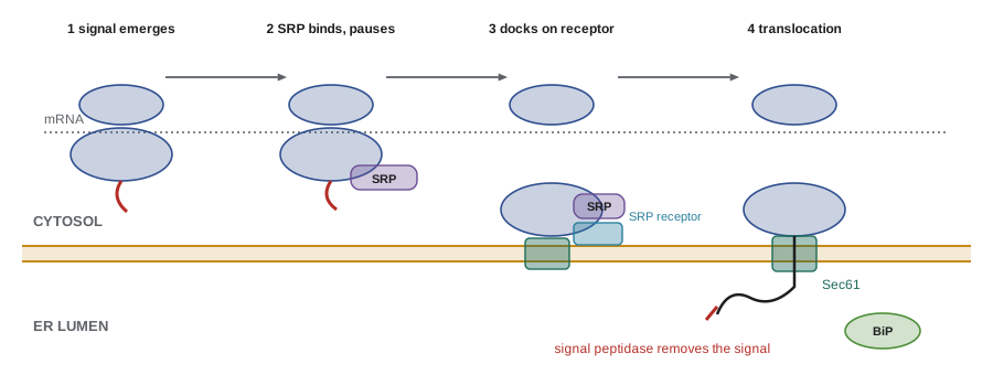
The Sec61 channel is an hourglass-shaped pore built from a ring of transmembrane helices, with a lateral gate that can open sideways into the lipid bilayer. A short plug blocks the channel when it is idle, keeping the ER’s ions and small molecules inside. When a hydrophobic segment of the growing chain passes through, it can slip sideways through the lateral gate into the membrane, which is how integral membrane proteins are inserted.
Some proteins are imported after they have been fully made, a process called post-translational translocation. In yeast this is common; the Sec61 channel works with additional proteins, and BiP, a chaperone of the Hsp70 family in the lumen, binds the incoming chain and acts as a ratchet, preventing it from sliding back. Very short proteins and tail-anchored proteins, which have a single hydrophobic segment at their extreme C-terminus, cannot use SRP because the signal emerges only after translation ends; they are captured by the GET pathway (TRC pathway in mammals), in which a cytosolic ATPase delivers them to a dedicated insertion complex.
How membrane proteins get their orientation
Integral membrane proteins have a defined orientation, or topology: each part of the chain is either on the cytosolic side, within the membrane, or on the luminal side, and this orientation is preserved as the protein travels on. The luminal side of the ER becomes the outside of the plasma membrane, because vesicle budding and fusion preserve membrane orientation. Topology is set during insertion by the arrangement of hydrophobic segments and charged residues.
| Type | How it is inserted | Final orientation | Example |
|---|---|---|---|
| Type I single-pass | Cleaved N-terminal signal peptide; an internal stop-transfer sequence halts translocation | N-terminus in lumen (outside cell) | LDL receptor, insulin receptor |
| Type II single-pass | Internal uncleaved signal-anchor sequence; N-terminus stays in cytosol | C-terminus in lumen | Transferrin receptor, many Golgi enzymes |
| Type III single-pass | Internal signal-anchor in the opposite orientation | N-terminus in lumen, no cleaved signal | Some cytochrome P450 enzymes |
| Tail-anchored | C-terminal hydrophobic segment inserted after translation (GET/TRC pathway) | N-terminus in cytosol | SNARE proteins, some mitochondrial outer membrane proteins |
| Multipass | Alternating start- and stop-transfer segments | Several crossings | G-protein-coupled receptors, transporters |
An empirical rule predicts much of this behavior: the positive-inside rule. Segments flanking a transmembrane helix that are rich in lysine and arginine tend to stay on the cytosolic side. Experiments that move positive charges from one flank to the other can flip a protein’s orientation. Together with hydrophobicity scans, the rule allows topology to be predicted from sequence.
# Predict transmembrane helices and orientation for an illustrative multipass protein.
KD = {"A": 1.8, "R": -4.5, "N": -3.5, "D": -3.5, "C": 2.5, "Q": -3.5, "E": -3.5, "G": -0.4,
      "H": -3.2, "I": 4.5, "L": 3.8, "K": -3.9, "M": 1.9, "F": 2.8, "P": -1.6, "S": -0.8,
      "T": -0.7, "W": -0.9, "Y": -1.3, "V": 4.2}
# Illustrative sequence with three hydrophobic segments (not a real protein)
seq = ("MSKRKSPEQ" "LLVALIAGFLVVLLAAI" "GNDTSEPGSWT" "FAVLIGLWLVSAFVILV"
       "KRKRGKSPAQ" "ILAVFLGLILAWVLLAV" "DNGSTEEQ")
w, cut = 17, 1.6
scores = [sum(KD[a] for a in seq[i:i+w]) / w for i in range(len(seq) - w + 1)]

# group consecutive high-scoring windows into segments
segs, i = [], 0
while i < len(scores):
    if scores[i] > cut:
        j = i
        while j + 1 < len(scores) and scores[j + 1] > cut: j += 1
        best = max(range(i, j + 1), key=scores.__getitem__)
        segs.append(best); i = j + 1
    else:
        i += 1

def pos_charge(s): return sum(s.count(x) for x in "KR")
print(f"{len(segs)} predicted transmembrane segments")
for k, s in enumerate(segs, 1):
    print(f"  TM{k}: residues {s+1}-{s+w}  {seq[s:s+w]}  mean hydropathy {scores[s]:.2f}")
first = segs[0]
n_side = pos_charge(seq[max(0, first-15):first])
c_side = pos_charge(seq[first+w:first+w+15])
print(f"K+R in 15 residues before TM1: {n_side}; after TM1: {c_side}")
print("positive-inside rule: N-terminus in the", "cytosol" if n_side > c_side else "lumen")3 predicted transmembrane segments TM1: residues 10-26 LLVALIAGFLVVLLAAI mean hydropathy 3.18 TM2: residues 38-54 FAVLIGLWLVSAFVILV mean hydropathy 2.83 TM3: residues 65-81 ILAVFLGLILAWVLLAV mean hydropathy 3.02 K+R in 15 residues before TM1: 3; after TM1: 0 positive-inside rule: N-terminus in the cytosol
The scan finds three hydrophobic segments, each long enough to span the bilayer as an α helix (about 20 residues cross the 3-nm hydrophobic core), and the basic residues before the first helix place the N-terminus in the cytosol. With three crossings, the C-terminus must then be in the lumen. Modern prediction programs combine such rules with machine learning and structural models, and are right for most proteins, but experiments such as glycosylation-site mapping, described below, are used to confirm them.
N-linked glycosylation
Most proteins made in the ER are glycoproteins. As a chain enters the lumen, an enzyme complex associated with the translocon, oligosaccharyltransferase, transfers a preassembled oligosaccharide of 14 sugars (two N-acetylglucosamine, nine mannose and three glucose residues, written Glc₃Man₉GlcNAc₂) from a lipid carrier, dolichol phosphate, onto the side chain of an asparagine. Only asparagines in the sequence Asn–X–Ser or Asn–X–Thr, where X is any amino acid except proline, can receive it; this motif is called a sequon. Not every sequon is used, but no N-linked sugar is added elsewhere. Because glycosylation happens only in the lumen, a sequon that becomes glycosylated must lie on the luminal side, which is why adding or removing sequons is a standard way to test topology.
import re
# Find N-glycosylation sequons (N-X-S/T, X not P) in an illustrative luminal domain.
seq = "QTNVSFKDLPNGTWAENQSPLNPTGHNETLSAQNISVYNPSEDKRNVT"
sequon = re.compile(r"(?=(N[^P][ST]))")
sites = [(m.start() + 1, m.group(1)) for m in sequon.finditer(seq)]
print("sequence:", seq)
print("sequons found:", sites)
blocked = [(m.start() + 1, seq[m.start():m.start()+3]) for m in re.finditer(r"(?=NP[ST])", seq)]
print("N-P-S/T motifs (not glycosylated):", blocked)
# each N-glycan of a complex type adds roughly 2 to 3 kDa
print(f"approximate added mass if all sites are used: {len(sites)*2.5:.1f} kDa")sequence: QTNVSFKDLPNGTWAENQSPLNPTGHNETLSAQNISVYNPSEDKRNVT sequons found: [(3, 'NVS'), (11, 'NGT'), (17, 'NQS'), (27, 'NET'), (34, 'NIS'), (46, 'NVT')] N-P-S/T motifs (not glycosylated): [(22, 'NPT'), (39, 'NPS')] approximate added mass if all sites are used: 15.0 kDa
The added sugars do several jobs. They make proteins more soluble, protect them from proteases, mark their folding state for quality control, and after trimming and modification in the Golgi (lesson 4) form part of the recognition signals on the cell surface, such as the ABO blood group antigens. Inherited defects in the enzymes that build or trim N-glycans cause a group of rare disorders called congenital disorders of glycosylation, which affect many organs.
Folding and quality control
The ER lumen is an oxidizing environment, unlike the cytosol, so disulfide bonds can form. Protein disulfide isomerase (PDI) catalyzes their formation and rearranges incorrect pairs until the right ones are found. BiP binds exposed hydrophobic stretches of unfolded chains and releases them in an ATP-driven cycle. A dedicated system, the calnexin–calreticulin cycle, uses the glycan itself as a folding tag.
Immediately after transfer, two of the three glucose residues on the glycan are trimmed. The lectin chaperones calnexin (membrane-bound) and calreticulin (soluble) bind proteins carrying a single glucose and hold them while they fold. When the last glucose is removed, the protein is released. If it has folded correctly, it can leave the ER. If it is still misfolded, an enzyme called UGGT, which recognizes exposed hydrophobic patches, adds a glucose back, and the protein rebinds calnexin for another try. Proteins that fail repeatedly have mannose residues trimmed, which marks them for destruction.
Terminally misfolded proteins are removed by ER-associated degradation (ERAD). They are recognized, pulled back across the ER membrane into the cytosol through channels formed by ubiquitin ligase complexes, ubiquitinated, extracted by the ATPase p97 (also called VCP) and degraded by the proteasome. The common cystic fibrosis mutation ΔF508 (BIOL-201, BIOL-207) produces a CFTR protein that is mostly caught by this quality control and destroyed even though it could partly work; corrector drugs help it escape.
Worked problem
How strict is ER quality control?
Suppose that each round of the calnexin cycle gives a newly made protein a 40 percent chance of folding correctly, and after five unsuccessful rounds the protein is sent to ERAD. What fraction of molecules folds, and what fraction is degraded? A mutation lowers the chance per round to 8 percent; recalculate.
Show solution
- Probability of failing all five rounds = (1 − p)⁵.
- Normal protein: 0.6⁵ = 0.078, so about 92 percent fold and 8 percent are degraded.
- Mutant protein: 0.92⁵ = 0.66, so only about 34 percent fold and 66 percent are degraded.
- A modest decrease in folding efficiency per round becomes a large loss of functional protein after quality control.
Answer: About 92 percent of the normal protein but only about 34 percent of the mutant protein escapes degradation (illustrative numbers).
The unfolded protein response
When unfolded proteins accumulate in the ER, because of a surge in secretory demand, a mutant protein, low glucose or drugs, cells activate the unfolded protein response (UPR). Three sensors in the ER membrane detect the problem, each normally held inactive by BiP; as unfolded proteins pull BiP away, the sensors are released.
| Sensor | Activation | Main output |
|---|---|---|
| IRE1 | Oligomerizes; its RNase domain splices XBP1 mRNA in the cytosol | XBP1s transcription factor increases chaperones and ERAD components |
| PERK | A kinase that phosphorylates the translation factor eIF2α | General translation slows, reducing the load; ATF4 is still translated and activates stress genes |
| ATF6 | Moves to the Golgi, where proteases release its cytosolic domain | ATF6 fragment enters the nucleus and induces chaperones and lipid synthesis |
Together these responses reduce the influx of new proteins, increase folding capacity and step up degradation. The splicing of XBP1 mRNA by IRE1 is unusual: it happens in the cytoplasm, not on the spliceosome. If stress cannot be relieved, prolonged signaling, especially through PERK and its downstream factor CHOP, triggers apoptosis (lesson 12). The UPR is especially important for professional secretory cells: pancreatic β cells, which make insulin, and plasma cells, which can each secrete thousands of antibody molecules per second. Chronic ER stress contributes to diabetes, neurodegeneration and some cancers, and drugs that modulate UPR branches are under investigation.
The ER makes lipids
Most membrane phospholipids are synthesized by enzymes on the cytosolic face of the ER membrane, using fatty acids and other precursors from the cytosol. New lipids are therefore added only to the cytosolic leaflet. Proteins called scramblases move phospholipids between leaflets in the ER, keeping the two halves balanced; in the plasma membrane, by contrast, ATP-driven flippases keep phosphatidylserine on the cytosolic side, and its appearance on the outer surface is a signal of apoptosis (lesson 12). The ER also makes cholesterol and ceramide, the precursor of sphingolipids, which are completed in the Golgi.
Lipids reach other organelles in two ways: in transport vesicles, and by direct transfer at membrane contact sites, places where the ER comes within 10 to 30 nm of another organelle. Lipid-transfer proteins bridge the gap, and at ER–mitochondria contacts calcium flows directly into mitochondria. Mitochondria and peroxisomes, which are not connected to the secretory pathway by vesicles, depend heavily on such contacts for their lipids.
Calcium storage
The ER lumen holds calcium at roughly 0.1 to 1 mM, compared with about 0.1 µM in the resting cytosol, a gradient of up to 10,000-fold maintained by SERCA pumps (sarco/endoplasmic reticulum Ca²⁺-ATPases). When a hormone signal produces inositol trisphosphate (IP₃, lesson 11), IP₃ receptors in the ER membrane open and calcium floods into the cytosol. In skeletal muscle, an action potential opens ryanodine receptors in the sarcoplasmic reticulum, releasing the calcium that triggers contraction (lesson 8); mutations in the skeletal muscle ryanodine receptor cause malignant hyperthermia, a dangerous reaction to some anesthetics.
Worked problem
Calcium release and cytosolic concentration
An ER compartment occupies 10 percent of a cell’s volume and contains free calcium at 0.5 mM. The cytosol is 50 percent of the cell volume. If 2 percent of the ER calcium is released and none were buffered, by how much would the cytosolic concentration rise?
Show solution
- Calcium released per liter of cell = 0.10 × 0.5 mM × 0.02 = 0.001 mM = 1 µM (expressed per cell volume).
- Spread over a cytosol of 0.5 of the cell volume: 1 µM / 0.5 = 2 µM.
- Resting cytosolic calcium is about 0.1 µM, so this is a 20-fold rise.
- In reality most released calcium is buffered by proteins, so free calcium rises less, typically to about 1 µM.
Answer: Without buffering, a rise of about 2 µM, a 20-fold increase over resting levels; buffering reduces the free rise.
Common mistakes
MistakeRibosomes are permanently attached to the rough ER.
InsteadRibosomes attach only while translating proteins with ER signals, then return to the cytosol.
MistakeProteins are pushed through the translocon by ATP hydrolysis.
InsteadIn co-translational import, ribosome synthesis drives the chain through; GTP hydrolysis by SRP and its receptor controls targeting.
MistakeAny asparagine can receive an N-linked glycan.
InsteadOnly Asn in Asn–X–Ser/Thr sequons where X is not proline.
MistakeMisfolded proteins are degraded inside the ER lumen.
InsteadERAD returns them to the cytosol for ubiquitination and degradation by the proteasome.
MistakeThe unfolded protein response always protects the cell.
InsteadIt first restores balance, but prolonged stress triggers apoptosis.
Summary and key terms
- The ER is a continuous membrane network with rough (ribosome-studded) and smooth regions.
- SRP, its receptor and the Sec61 translocon carry out co-translational import; tail-anchored proteins use the GET/TRC pathway.
- Stop-transfer and signal-anchor sequences and the positive-inside rule set membrane protein topology.
- Oligosaccharyltransferase adds Glc₃Man₉GlcNAc₂ to Asn in N–X–S/T sequons.
- PDI, BiP and the calnexin–calreticulin cycle fold proteins; ERAD destroys those that fail.
- The unfolded protein response acts through IRE1, PERK and ATF6; prolonged stress triggers apoptosis.
- The ER makes lipids, transfers them at contact sites and stores calcium released by IP₃ and ryanodine receptors.
- rough ER
- ER studded with ribosomes making secretory and membrane proteins.
- Sec61 translocon
- The protein-conducting channel through which chains enter or insert into the ER membrane.
- stop-transfer sequence
- A hydrophobic segment that halts translocation and becomes a membrane-spanning helix.
- signal-anchor sequence
- An internal uncleaved hydrophobic segment that both targets and anchors a protein.
- positive-inside rule
- Lysine- and arginine-rich flanks of transmembrane helices tend to face the cytosol.
- sequon
- The Asn–X–Ser/Thr motif (X not Pro) required for N-linked glycosylation.
- calnexin
- A membrane lectin chaperone that binds monoglucosylated glycoproteins during folding.
- ERAD
- ER-associated degradation, the retrotranslocation and proteasomal destruction of misfolded ER proteins.
- unfolded protein response
- Signaling from ER stress sensors that restores folding balance or triggers apoptosis.
- membrane contact site
- A region where two organelle membranes come close and exchange lipids or ions directly.
Practice problems
Work each problem on paper first, then reveal the answer to check.
Why do pancreatic acinar cells have abundant rough ER?
Show answer
They secrete large amounts of digestive enzymes, all made on ER-bound ribosomes.
What provides the energy for moving a chain through the translocon during co-translational import?
Show answer
Ongoing protein synthesis by the ribosome sitting on the channel.
A single-pass protein has a cleaved signal peptide and an internal stop-transfer segment. Which end faces the lumen?
Show answer
The N-terminus (type I protein).
How do tail-anchored proteins reach the ER membrane?
Show answer
Post-translationally, through the GET/TRC pathway, because their signal emerges only after translation ends.
Which of these can be glycosylated: NPT, NAS, NGT?
Show answer
NAS and NGT; NPT cannot because proline is in the X position.
What does UGGT do?
Show answer
It adds a glucose back to misfolded glycoproteins so they rebind calnexin for another folding attempt.
Name the three UPR sensors.
Show answer
IRE1, PERK and ATF6.
How does PERK reduce ER load?
Show answer
It phosphorylates eIF2α, slowing general translation.
Where are most new phospholipids added, and how do they reach the other leaflet?
Show answer
To the cytosolic leaflet of the ER; scramblases move them across.
If folding succeeds with probability 0.3 per round and five rounds are allowed, what fraction folds?
Show answer
1 − 0.7⁵ ≈ 0.83.
Lesson quiz · 5 questions
Score 60% or more to complete this lesson.
Create a free account to save your progress, take quizzes and earn a certificate.