
HSE-394 · Lesson 9 of 12 · 22 min
Ionizing radiation: principles of protection
▶ Video coming soon A video version of this lesson is on the way.
Ionizing radiation is one of the most useful tools in medicine. X-rays show broken bones and pneumonia, CT scans find bleeding in the brain, fluoroscopy guides catheters into the heart, and radioactive drugs reveal how organs work and treat some cancers. It is also one of the best understood hazards in the workplace, with a century of research behind its limits and controls. This lesson explains what ionizing radiation is, how dose is measured, what radiation does to the body, the three basic protections of time, distance and shielding, the dose limits, and how workers' doses are measured. The next lesson applies these principles in imaging and nuclear medicine.
What ionizing radiation is
Radiation is energy travelling as waves or particles. Ionizing radiation carries enough energy to knock electrons out of atoms, creating charged particles called ions. When this happens in living tissue, it can break chemical bonds, including those in DNA. Non-ionizing radiation, such as radio waves, microwaves, visible light and the fields used in MRI, does not have enough energy to do this; its hazards, covered in Lesson 11, are different.
The types that matter in healthcare are:
- X-rays: produced by a machine when a high voltage accelerates electrons into a metal target. They exist only while the machine is switched on and producing a beam. When the beam stops, there is no radiation left.
- Gamma rays: similar to X-rays but given off by radioactive atoms as they decay, such as technetium-99m used in nuclear medicine. A radioactive source keeps emitting until it has decayed.
- Beta particles: fast electrons given off by some radioactive materials, such as iodine-131 and fluorine-18 (which gives off positrons, the positive version). They travel a short distance in tissue but can burn skin if spilled on it.
- Alpha particles: heavy particles that cannot pass through skin but are very damaging if the material is swallowed or breathed in. They are used in a few cancer treatments.
Measuring dose
Two units matter for workers.
- The gray (Gy) measures absorbed dose: the energy deposited in tissue. A milligray (mGy) is a thousandth of a gray.
- The sievert (Sv) measures effective dose: the absorbed dose adjusted for the type of radiation and the sensitivity of the tissues exposed, so that doses to different parts of the body can be compared as a whole-body risk. For X-rays and gamma rays, 1 mGy to the whole body equals about 1 mSv. Occupational doses are usually recorded in millisieverts (mSv) or microsieverts.
In the United States you will also see older units: the rad for absorbed dose (1 Gy = 100 rad) and the rem for effective dose (1 Sv = 100 rem, so 1 mSv = 100 millirem). Canadian regulations use SI units only.
To put numbers in context: everyone receives radiation from natural sources, including radon in homes, cosmic rays and radioactive elements in rocks, food and our own bodies. Natural background averages around 2 to 3 mSv a year in North America, varying with where people live. A chest X-ray gives the patient about 0.02 to 0.1 mSv. A CT scan of the abdomen gives several mSv. Most hospital workers who are monitored receive less than 1 mSv a year above background, and many receive nothing measurable. A smaller group, especially those who work close to patients during fluoroscopy and some nuclear medicine staff, can receive more, which is why their doses are tracked closely.
What radiation does to the body
Radiation effects fall into two groups.
- Deterministic effects (also called tissue reactions) happen only above a threshold dose, and get worse as the dose increases. Examples are skin reddening and burns, hair loss, cataracts in the lens of the eye, and, at very high doses, radiation sickness. Below the threshold they do not occur. In healthcare, the main deterministic risks to workers are cataracts in staff who work near fluoroscopy beams for years without eye protection, and skin injury to hands placed in the beam.
- Stochastic effects are effects of chance: cancer, and in theory inherited effects. Their probability rises with dose, but their severity does not. For radiation protection, regulators assume that any dose, however small, carries some added cancer risk, in proportion to the dose. This assumption, called the linear no-threshold model, is cautious, and it is the basis of dose limits and of ALARA.
The unborn child is more sensitive to radiation than an adult, especially in the first months of pregnancy, which is why lower limits apply once a worker has declared a pregnancy.
ALARA
ALARA stands for "as low as reasonably achievable". It means keeping doses as far below the limits as is reasonably possible, taking costs and practicality into account. A worker whose dose is well under the limit is not finished with protection; the question is always whether the dose could reasonably be lower. Every licence and registration for radiation work requires an ALARA program.
Time, distance and shielding
Three simple principles reduce dose from any source of external radiation.
- Time: dose is proportional to time spent near the source. Halving the time halves the dose. Plan the task, have supplies ready, and do not linger near a patient during an X-ray exposure or near a radioactive patient longer than care requires.
- Distance: radiation spreads out as it travels, so its intensity falls with the square of the distance from a small source. This is the inverse square law. Doubling the distance cuts the dose to a quarter; tripling it cuts the dose to a ninth. Distance is the cheapest and most powerful protection.
- Shielding: dense materials absorb radiation. Lead aprons, thyroid collars, leaded glasses, ceiling-suspended shields, mobile shields and lead-lined walls are all forms of shielding. A typical lead apron, rated at 0.25 to 0.5 mm of lead equivalent, absorbs most of the scattered X-rays that reach it, but it does nothing for the parts of the body it does not cover.
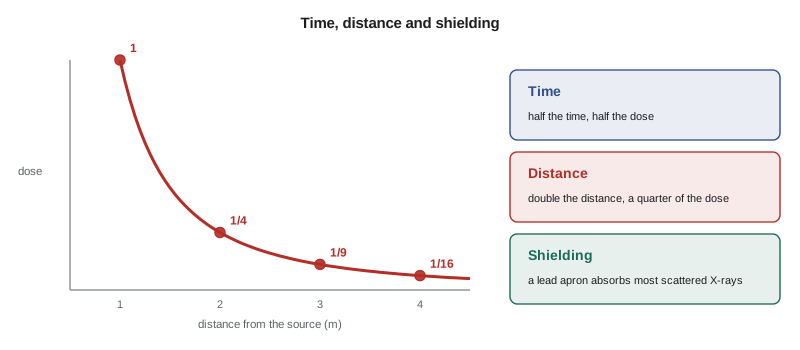
The arithmetic of distance is worth practising, because it tells staff how far to step back. The radiation safety officer at Northbridge, Dr. Helen Ward, uses a short calculation in her training sessions to show nurses how their dose changes when they stand in different places during a portable X-ray.
# Inverse square law: dose to a nurse near a patient during portable X-rays
# Scattered dose rate measured at 1 m from the patient during one exposure
dose_at_1m = 1.0   # microsieverts per exposure (illustrative)

def dose(distance_m, exposures=1, shield_factor=1.0):
    return dose_at_1m * exposures * shield_factor / distance_m ** 2

print(f"{'where the nurse stands':34}{'per exposure':>13}{'per year':>10}")
places = [("0.5 m, holding the patient", 0.5, 1.0),
          ("1 m, beside the bed", 1.0, 1.0),
          ("2 m, at the foot of the bed", 2.0, 1.0),
          ("3 m, in the doorway", 3.0, 1.0),
          ("0.5 m, holding, with apron", 0.5, 0.1)]
for label, d, s in places:
    per = dose(d, 1, s)
    year = dose(d, 300, s)    # 300 portable films a year on a busy unit
print(f"{label:34}{per:10.2f} uSv{year:8.0f} uSv")where the nurse stands per exposure per year 0.5 m, holding the patient 4.00 uSv 1200 uSv 1 m, beside the bed 1.00 uSv 300 uSv 2 m, at the foot of the bed 0.25 uSv 75 uSv 3 m, in the doorway 0.11 uSv 33 uSv 0.5 m, holding, with apron 0.40 uSv 120 uSv
The numbers are illustrative, but the pattern is real. Standing 2 m (6 ft) away cuts the dose by four compared with standing at 1 m, and by sixteen compared with holding the patient at half a metre. Over a year of portable X-rays, the difference between stepping back and staying close is large. Wearing an apron helps, but only for the parts of the body it covers, and it is much better to step away than to stay close with an apron on. When a patient must be held, the holder should be someone who is not regularly exposed, such as a family member given an apron, never the same nurse every time, and never a pregnant worker.
Dose limits
In Canada, the Radiation Protection Regulations under the Nuclear Safety and Control Act set dose limits for work with nuclear substances, enforced by the CNSC. A worker whose job involves a reasonable chance of significant exposure, such as nuclear medicine staff, is designated a nuclear energy worker (NEW) and informed in writing. The limits for a NEW are 50 mSv in any one year and 100 mSv over a five-year period, and, since 2021, 50 mSv a year to the lens of the eye. A pregnant NEW who informs the employer in writing has a limit of 4 mSv for the balance of the pregnancy. Workers who are not NEWs, and the public, have a limit of 1 mSv a year. For X-ray equipment, Ontario's X-ray Safety regulation under the OHSA sets dose limits for X-ray workers and lower limits for other workers, including a lower limit for pregnant workers, and requires the employer to protect workers who are not X-ray workers.
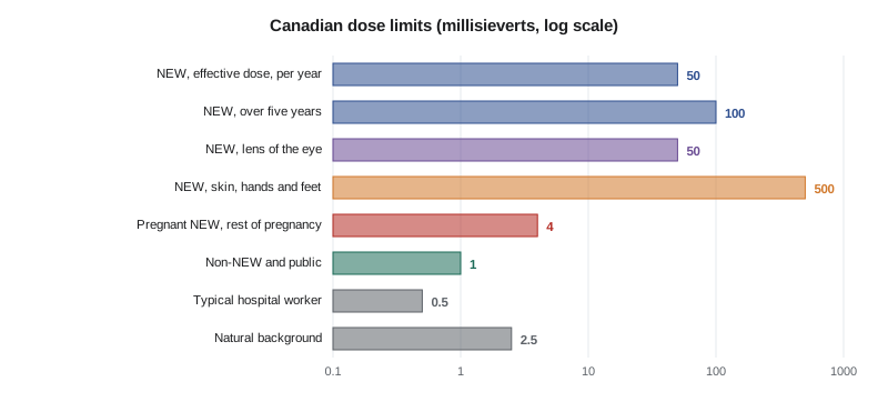
Limits are a ceiling, not a target. A worker approaching a limit is a sign that something in the work, the equipment or the protection needs to change. Most hospitals set lower investigation levels, at which the radiation safety officer reviews a worker's dose and practices, long before any limit is in sight.
Measuring workers' doses
Workers who could receive a significant dose wear a personal dosimeter, usually a small badge containing a material that records the radiation that passes through it. Badges are sent to a dosimetry service and read monthly or every three months, and the results become part of the worker's permanent dose record.
- The main badge is worn on the trunk, between the waist and the neck. For staff who wear lead aprons in fluoroscopy, the badge is worn at the collar, outside the apron, so that it estimates the dose to the head and eyes; some programs add a second badge under the apron.
- Ring dosimeters are worn on the hand most likely to be exposed by staff who handle radioactive drugs or work with their hands near the beam.
- Electronic dosimeters that show the dose in real time are used in some interventional and nuclear medicine areas.
- Badges must be worn only by the person they belong to, stored away from radiation when not worn, and never left on an apron hanging in the X-ray room.
NEWs' doses are reported to Health Canada's National Dose Registry, which keeps a lifetime record of occupational doses for monitored workers across Canada.
Worked problem
The badge on the apron rack
Owen Fraser, a medical radiation technologist at Northbridge, receives a dosimetry report showing his badge recorded 3 mSv in one month, far above his usual readings of almost nothing. He remembers that he left his apron, with the badge clipped to the collar, hanging in the fluoroscopy room for most of that month. What should happen?
Show solution
- Owen should tell Dr. Ward, the radiation safety officer, right away, explaining how the badge was stored.
- Dr. Ward investigates: she checks Owen's work schedule and the doses of colleagues who did similar work, looks at the apron rack's position relative to the fluoroscopy unit, and may place a test badge there.
- If the investigation shows the badge was exposed while not being worn, the dose can be corrected in the record with a documented explanation, as dosimetry rules allow.
- The apron rack is moved out of the room or behind shielding, and staff are reminded to store badges in the designated badge rack outside the room.
Answer: Report it to the radiation safety officer, who investigates and, if the dose was not received by Owen, corrects the record with documentation; then fix the storage practice.
Common mistakes
MistakeThinking that an X-ray room stays radioactive after an exam.
InsteadX-rays stop when the machine stops; only radioactive materials keep emitting.
MistakeStaying close to the patient with a lead apron instead of stepping back.
InsteadStep back first; distance is the most powerful protection.
MistakeTreating the dose limit as a target.
InsteadKeep doses as low as reasonably achievable, well below the limits.
MistakeLeaving a dosimeter on an apron in the X-ray room.
InsteadStore badges outside the room, away from radiation.
MistakeAsking the same nurse to hold patients for X-rays every time.
InsteadUse holding devices, or rotate non-regular holders given aprons; never a pregnant worker.
Summary and key terms
- Ionizing radiation can break chemical bonds in tissue, including DNA.
- X-rays exist only while the machine is on; radioactive materials keep emitting as they decay.
- Absorbed dose is measured in grays and effective dose in sieverts; worker doses are usually in millisieverts.
- Deterministic effects such as cataracts have thresholds; stochastic effects such as cancer are assumed to rise with any dose.
- Time, distance and shielding reduce dose; doubling distance cuts dose to a quarter.
- Canadian limits: NEW 50 mSv a year and 100 mSv over five years, lens 50 mSv, pregnant NEW 4 mSv, others 1 mSv.
- Personal dosimeters record worker doses; they must be worn correctly and stored away from radiation.
- Ionizing radiation
- Radiation with enough energy to remove electrons from atoms.
- Gray (Gy)
- The unit of absorbed dose.
- Sievert (Sv)
- The unit of effective dose, used to compare risk.
- Deterministic effect
- A radiation effect that occurs only above a threshold dose, such as a cataract or skin burn.
- Stochastic effect
- A radiation effect, such as cancer, whose probability rises with dose.
- ALARA
- As low as reasonably achievable: keeping doses as far below limits as reasonably possible.
- Inverse square law
- Radiation intensity from a small source falls with the square of the distance.
- Dosimeter
- A device worn by a worker to measure radiation dose.
Practice problems
Work each problem on paper first, then reveal the answer to check.
What is the difference between X-rays and gamma rays?
Show answer
X-rays are made by a machine and stop when it is switched off; gamma rays come from radioactive atoms that keep decaying.
If a nurse moves from 1 m to 3 m from a patient during an X-ray, by how much does her dose fall?
Show answer
To a ninth of the dose at 1 m.
Name one deterministic and one stochastic effect of radiation.
Show answer
Deterministic: cataract, skin burn or hair loss; stochastic: cancer.
What is the dose limit for a pregnant NEW who has informed her employer?
Show answer
4 mSv for the balance of the pregnancy.
Where should a fluoroscopy worker wear the main dosimeter, and why?
Show answer
At the collar outside the apron, to estimate the dose to the head and eyes.
Why did Owen's badge show a high reading?
Show answer
It was left on an apron hanging in the fluoroscopy room, recording dose he did not receive.
Lesson quiz · 5 questions
Score 60% or more to complete this lesson.
Create a free account to save your progress, take quizzes and earn a certificate.