
CHE-260 · Lesson 11 of 12 · 22 min
Mass spectrometry and hyphenated methods
A customer of Kingfisher Bioenergy reports a faint off-odor in a shipment of fuel ethanol. The gas chromatograph shows a small unknown peak between isobutanol and isoamyl alcohol, at a retention time that matches nothing in the lab's standards. Retention time can suggest an identity, but it cannot prove one. To identify the compound, the lab sends the sample to its gas chromatograph-mass spectrometer, which records a mass spectrum of every peak as it leaves the column. Within an hour, a library search names the unknown, and the spectrum's isotope pattern and fragments confirm it. Mass spectrometry is the most powerful identification tool in the analytical lab, and coupled to chromatography it is also one of the most sensitive ways to measure trace compounds.
What a mass spectrometer does
A mass spectrometer does three things. It turns molecules into gas-phase ions, separates the ions according to their mass-to-charge ratio, m/z, and counts them. The output is a mass spectrum, a plot of ion abundance against m/z. Most ions carry a single charge, so m/z is usually the ion's mass in daltons (atomic mass units). Because separation requires ions to travel without colliding with gas molecules, the analyzer operates under high vacuum, about \(10^{-5}\) Pa or lower.
Ionization
Electron ionization (EI) is the classic method for gas chromatography. Molecules leaving the column pass through a beam of electrons accelerated to 70 eV, which knock an electron out of each molecule to form a radical cation, the molecular ion \(M^{+\bullet}\). The 70 eV electrons deliver far more energy than is needed, so many molecular ions break apart into fragment ions in patterns that are characteristic and highly reproducible. Because every instrument uses 70 eV, EI spectra can be compared with large reference libraries containing hundreds of thousands of compounds. The drawback is that for some compounds the molecular ion fragments so completely that it barely appears.
Chemical ionization uses a reagent gas such as methane or ammonia, which is ionized first and then transfers a proton to the analyte gently, giving a strong \([M+H]^+\) ion and little fragmentation, which helps confirm molecular mass. For liquid chromatography, electrospray ionization (ESI) is dominant. The column effluent is sprayed from a fine needle held at several kilovolts, forming charged droplets that shrink as solvent evaporates until ions are released into the gas phase. ESI is gentle enough to ionize sugars, mycotoxins, proteins and other fragile molecules, usually as \([M+H]^+\) or \([M-H]^-\), and it can put many charges on large molecules such as proteins.
Mass analyzers
| Analyzer | How it separates ions | Strengths |
|---|---|---|
| Quadrupole | Oscillating electric fields on four rods let only one m/z through at a time | Robust, inexpensive, excellent for quantitation |
| Triple quadrupole | Two quadrupoles separated by a collision cell | Selects a parent ion and a fragment for highly selective trace quantitation |
| Ion trap | Traps ions in electric fields and ejects them by m/z | Compact, can fragment ions repeatedly |
| Time of flight (TOF) | Ions given the same energy reach the detector at times depending on m/z | Fast, high mass accuracy, wide mass range |
| Orbital trap | Ions orbit an electrode at frequencies depending on m/z | Very high resolving power and mass accuracy |
Resolving power describes how well an analyzer separates ions of nearly equal mass. A quadrupole has unit resolution, distinguishing m/z 46 from 47 but not 46.04 from 46.07. High-resolution instruments such as TOF and orbital traps can measure m/z to a few parts per million, which is enough to tell which elemental formula an ion has. Carbon monoxide and nitrogen both have a nominal mass of 28, but their exact masses are 27.9949 and 28.0061, easily distinguished at high resolution.
Reading a mass spectrum
The most intense peak in a spectrum is the base peak, set to 100 percent, and other peaks are scaled to it. The molecular ion, when present, gives the molar mass. Fragments reveal structure, because bonds break in predictable ways. Alcohols lose a hydrogen or alkyl group next to the oxygen to form stable oxygen-containing cations: ethanol (molar mass 46) shows a small molecular ion at m/z 46, a peak at 45 from loss of a hydrogen atom, and a base peak at 31 from \(\text{CH}_2\text{OH}^+\), formed when the methyl group breaks off. Ethyl acetate (88) shows a weak molecular ion at 88, fragments at 70 and 61, and a base peak at 43 from the acetyl ion \(\text{CH}_3\textKingfisher Bioenergy^+\). Losses of 15 (methyl), 18 (water) and 28 (ethene or carbon monoxide) are common and worth recognizing.
The nitrogen rule is a quick check: a molecule with an odd number of nitrogen atoms has an odd molar mass, and one with zero or an even number of nitrogen atoms has an even molar mass. A molecular ion at m/z 59 therefore suggests one nitrogen.
Isotope patterns
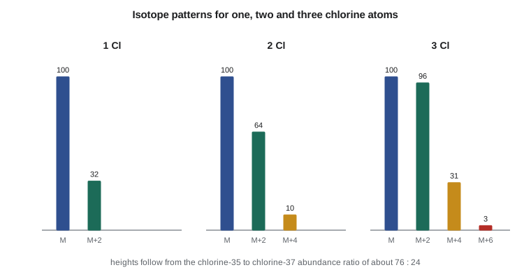
Most elements have more than one stable isotope, and their natural abundances leave fingerprints in mass spectra. About 1.07 percent of carbon is carbon-13, so a molecule with n carbon atoms has an M+1 peak of roughly \(1.1n\) percent of the molecular ion. That lets the analyst estimate the number of carbons: an M+1 peak of 8.7 percent suggests about eight carbons. Chlorine and bromine are even more distinctive. Chlorine is about 76 percent chlorine-35 and 24 percent chlorine-37, so one chlorine gives an M+2 peak about one-third the height of M; bromine's two isotopes are nearly equal, so one bromine gives M and M+2 peaks of almost equal height. Two or three halogens give patterns predicted by the binomial expansion.
Worked problem
Identifying the off-odor compound
The unknown peak's EI spectrum shows a molecular ion at m/z 102 with an M+1 peak of 5.6 percent, a base peak at 43 and significant peaks at 61 and 73. No M+2 peak is visible. The library suggests propyl acetate. Is the spectrum consistent with that?
Show solution
- Propyl acetate, \(\text{C}_5\text{H}_{10}\text{O}_2\), has molar mass 102, matching the molecular ion. Its even mass fits zero nitrogens.
- The M+1 peak of 5.6 percent suggests about \(5.6/1.1 \approx 5\) carbons, matching the formula; five carbons predict 5.4 percent, and the small extra comes from deuterium and oxygen-17.
- The base peak at 43 is the acetyl ion typical of acetates; 61 is protonated acetic acid, also typical of acetate esters; 73 is loss of an ethyl group (102 − 29) from the propyl chain.
- No M+2 peak rules out chlorine and bromine.
Answer: The spectrum is consistent with propyl acetate, a fruity-smelling ester. The lab confirms by running a pure standard to match both the retention time and the spectrum, then traces the source to a fermentation upset that raised the higher alcohols, which form esters during distillation and storage.
Measuring with mass spectrometry
Identification is only half the job. Once the lab knows the off-odor compound is propyl acetate, the customer wants to know how much is present, and the plant wants to know whether other shipments contain it. GC-MS quantitation works like any other chromatographic calibration, with standards and usually an internal standard, but it measures the area of one characteristic ion rather than the whole peak. A quantifier ion, chosen for its intensity and freedom from interference, gives the number. One or two qualifier ions confirm identity: their ratio to the quantifier must match the ratio in a pure standard within a set tolerance, typically 20 percent, or the result is flagged.
Mass spectrometers need regular care. The ion source gradually becomes coated with sample residues, which lowers sensitivity, and the instrument is tuned against a reference compound to check mass calibration and ion ratios before each sequence. The vacuum pumps, the GC column's bleed and the carrier gas purity all affect the background. Because a mass spectrometer is expensive and needs skilled operators, many plants keep only GC and HPLC instruments in-house and send identification work to a contract laboratory. Kingfisher Bioenergy bought its GC-MS after a year of recurring odor complaints made the contract lab's turnaround too slow.
Exact mass
High-resolution instruments report m/z to four or more decimal places. Comparing the measured mass with the theoretical exact mass of a proposed formula, calculated from the monoisotopic masses of the most abundant isotopes, gives the mass error in parts per million:
An error within about 5 ppm, together with a sensible isotope pattern, usually narrows a small molecule down to one or a few elemental formulas. The program below calculates carbon-13 M+1 ratios for three compounds found in fuel ethanol, the chlorine isotope patterns in the figure, and the mass error for protonated ethanol measured on a high-resolution instrument.
# Mass spectrometry: isotope patterns, exact mass error and fragment checks
from math import comb
# 1. M+1 peak from carbon-13 (natural abundance about 1.07 percent)
p13 = 0.0107
for name, nC in [('ethanol C2H6O', 2), ('isoamyl alcohol C5H12O', 5), ('ethyl hexanoate C8H16O2', 8)]:
    ratio = nC * p13 / (1 - p13) * 100
    print(f"{name:26s} M+1 / M = {ratio:.1f} %")

# 2. Chlorine pattern: Cl-35 75.8 %, Cl-37 24.2 %
def cl_pattern(n):
    p, q = 0.758, 0.242
    peaks = [comb(n, k) * p ** (n - k) * q ** k for k in range(n + 1)]
    top = max(peaks)
    return [round(x / top * 100) for x in peaks]
for n in (1, 2, 3):
    print(f"{n} Cl: M, M+2, ... = {cl_pattern(n)}")

# 3. Exact mass and ppm error for a high-resolution measurement
mono = {'C': 12.000000, 'H': 1.007825, 'O': 15.994915, 'N': 14.003074}
def mass(f): return sum(mono[e] * k for e, k in f.items())
ion = mass({'C': 2, 'H': 6, 'O': 1}) + 1.007276          # protonated ethanol [M+H]+
measured = 47.04913
print(f"theoretical [M+H]+ = {ion:.5f}, error = {(measured - ion) / ion * 1e6:.1f} ppm")ethanol C2H6O M+1 / M = 2.2 % isoamyl alcohol C5H12O M+1 / M = 5.4 % ethyl hexanoate C8H16O2 M+1 / M = 8.7 % 1 Cl: M, M+2, ... = [100, 32] 2 Cl: M, M+2, ... = [100, 64, 10] 3 Cl: M, M+2, ... = [100, 96, 31, 3] theoretical [M+H]+ = 47.04914, error = -0.2 ppm
Hyphenated methods
Coupling a separation to a spectroscopic detector creates a hyphenated method, and the combination is far more powerful than either alone. In gas chromatography-mass spectrometry (GC-MS), the column effluent flows directly into the ion source, and the instrument records a full spectrum several times per second. The total ion chromatogram looks like an ordinary chromatogram, but every point carries a spectrum, so each peak can be identified by library search. When the target compounds are known, the instrument can record only a few characteristic ions (selected ion monitoring), which raises sensitivity a hundredfold by spending all its time on the ions that matter.
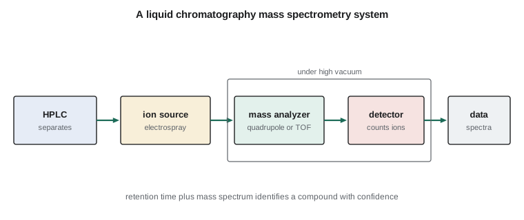
Liquid chromatography-tandem mass spectrometry (LC-MS/MS) on a triple quadrupole instrument is the standard for trace quantitation in complex samples. The first quadrupole selects the analyte's parent ion, the collision cell breaks it into fragments, and the third quadrupole passes only a chosen fragment. This multiple reaction monitoring is so selective that compounds can be measured at micrograms per kilogram in feed, food and environmental samples with little cleanup. It is how reference labs measure a dozen mycotoxins in distillers grains in one run, using isotopically labeled internal standards, versions of each toxin made with carbon-13, which behave identically in the column and ion source but appear at a different mass.
Other hyphenated methods include GC with infrared detection, LC with diode-array UV detection, which records a UV spectrum for every peak to check purity, and the ICP-MS coupled to liquid chromatography used to measure the different chemical forms of arsenic or selenium. In each case, one technique separates and the other identifies.
Common mistakes
MistakeAssuming the largest peak in a mass spectrum is the molecular ion.
InsteadThe largest peak is the base peak; the molecular ion may be small or absent in EI spectra.
MistakeAccepting a library match without checking it.
InsteadConfirm with a pure standard's retention time and spectrum, and check isotope patterns and the molecular ion.
MistakeUsing low-resolution data to assign an exact formula.
InsteadUnit-resolution masses fit many formulas; elemental formulas need high-resolution data within a few ppm.
MistakeForgetting matrix effects in electrospray LC-MS.
InsteadCo-eluting compounds can suppress ionization; use isotopically labeled internal standards or matrix-matched calibration.
Summary and key terms
- A mass spectrometer ionizes molecules, separates the ions by m/z under vacuum and counts them.
- Electron ionization at 70 eV gives reproducible fragmentation for library searching; electrospray ionizes fragile molecules for LC-MS.
- Quadrupoles are robust for quantitation; TOF and orbital traps give high resolution and exact mass.
- The molecular ion gives molar mass, fragments give structure, and the base peak is the most intense ion.
- Carbon-13 M+1 peaks estimate the number of carbons; chlorine and bromine give distinctive M+2 patterns.
- GC-MS identifies unknowns; LC-MS/MS with multiple reaction monitoring quantifies traces in complex samples.
- Mass-to-charge ratio
- The quantity by which a mass spectrometer separates ions, m/z.
- Electron ionization
- Ionization by a beam of 70 eV electrons, producing molecular and fragment ions.
- Electrospray ionization
- A gentle ionization method that sprays liquid from a charged needle to release ions from evaporating droplets.
- Molecular ion
- The ion formed from the intact molecule, which gives its molar mass.
- Base peak
- The most abundant ion in a mass spectrum, set to 100 percent.
- Resolving power
- The ability of a mass analyzer to separate ions of nearly equal m/z.
- Selected ion monitoring
- Recording only a few chosen ions to increase sensitivity for known targets.
- Multiple reaction monitoring
- Tandem MS quantitation that selects a parent ion and one of its fragments.
Practice problems
Work each problem on paper first, then reveal the answer to check.
Why do mass spectrometers operate under high vacuum?
Show answer
So ions can travel through the analyzer without colliding with gas molecules.
Ethanol's base peak is at m/z 31. What ion is it?
Show answer
\(\text{CH}_2\text{OH}^+\), formed by loss of a methyl group from the molecular ion.
An ion's M+1 peak is 4.4 percent of M. About how many carbons does it contain?
Show answer
About \(4.4/1.1 = 4\) carbons.
An ion shows M and M+2 peaks of nearly equal height. What element is likely present?
Show answer
One bromine atom.
A measured m/z is 103.07540 and the theoretical value is 103.07536. What is the error?
Show answer
\((0.00004/103.07536) \times 10^6 = 0.4\) ppm.
Why are isotopically labeled internal standards used in LC-MS/MS?
Show answer
They behave like the analyte in separation and ionization, so they correct for losses and ion suppression, but appear at a different mass.
Lesson quiz · 5 questions
Score 60% or more to complete this lesson.
Create a free account to save your progress, take quizzes and earn a certificate.